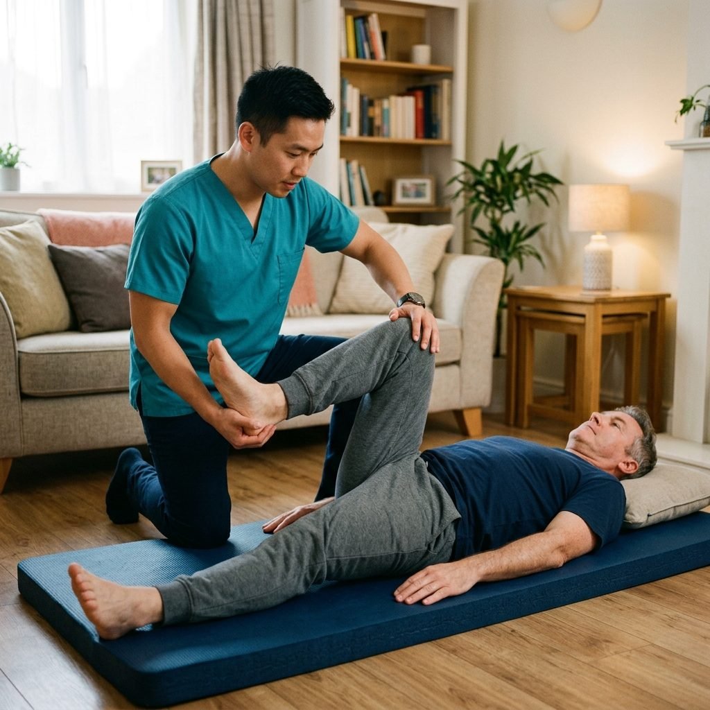
Sciatica is not a distinct medical diagnosis, but rather a clinical syndrome characterized by severe radiating pain along the path of the sciatic nerve. This nerve, the longest and thickest in the human body, branches from your lower back through your hips and buttocks and down the posterior aspect of each leg. Typically, sciatica affects only one side of your body. The condition usually arises when a herniated disc, a bone spur on the spine, or narrowing of the spinal canal (spinal stenosis) compresses part of the nerve. This mechanical compression is almost always accompanied by an intense local inflammatory response, leading to severe localized pain, numbness, tingling sensations, and sometimes muscular weakness in the affected leg. Although the pain associated with sciatica can be excruciating and profoundly impact daily life, the vast majority of cases resolve completely with conservative treatments such as specialized, evidence-based physical therapy. Severe cases involving significant neurological deficits, such as profound weakness or bowel and bladder dysfunction, are rare but require immediate surgical evaluation. Our comprehensive rehabilitation approach focuses on reducing nerve irritation, restoring normal spinal kinematics, and empowering patients with the precise tools needed to manage their spinal health independently over the long term.
Anatomy and Physiology of the Sciatic Nerve and Its Complex Pathway

The sciatic nerve is the largest and longest single nerve in the human body, serving as a major and vital neurological conduit between the central nervous system (spinal cord) and the lower extremities. It originates from the lumbosacral plexus, a complex network formed by the convergence of nerve roots from the lower lumbar spine (L4 and L5) and the sacrum (S1, S2, and S3). These individual nerve roots exit the protective spinal canal through narrow openings called intervertebral foramina, eventually joining and intertwining deep in the pelvic region to form the thick, continuous bundle of the sciatic nerve. This complex multi-root origin signifies that structural issues at various different levels of the lower spine—such as a herniated disc at L4-L5 or L5-S1—can all manifest clinically as sciatic pain, making precise neurological assessment essential to pinpoint the exact level of injury.
After its formation, the sciatic nerve exits the pelvis through the greater sciatic foramen, passing deep to the gluteus maximus muscle. Crucially, its pathway takes it in extremely close proximity to the piriformis muscle, a small but powerful external rotator of the hip. In the vast majority of individuals, the nerve travels just beneath the piriformis. However, significant anatomical variations exist where the nerve, or a major division of it, may actually pierce and travel directly through the piriformis muscle belly itself. This anatomical configuration predisposes these individuals to a distinct type of nerve entrapment known as 'piriformis syndrome,' where spasms or hypertrophy of the muscle physically strangle the nerve, perfectly mimicking the symptoms of a true spinal disc herniation. The nerve then descends down the posterior aspect of the thigh, providing essential motor innervation to the powerful hamstring muscle group.
Just above the posterior aspect of the knee (in the popliteal fossa), the sciatic nerve naturally bifurcates into two major, distinct branches: the tibial nerve and the common fibular (peroneal) nerve. These branches continue their descent to provide sensory and motor innervation to the entire lower leg, ankle, and foot. The physiological health of the sciatic nerve relies heavily on its microscopic anatomical integrity, which includes robust protective sheaths (epineurium, perineurium, and endoneurium) and a highly dedicated, intricate blood supply known as the vasa nervorum. When the nerve is subjected to sustained mechanical compression or excessive pathological tension, these delicate, microscopic blood vessels become compressed and compromised, leading to acute nerve ischemia (lack of oxygen). This state of ischemia, combined with the physical deformation of the neural fibers, severely impairs normal neurological conduction and generates continuous, agonizing ectopic pain signals. Restoring unhindered blood flow and alleviating this mechanical pressure are therefore the primary physiological goals of any effective sciatica rehabilitation program.
Pathophysiological Mechanisms of Sciatica and Nerve Root Compression
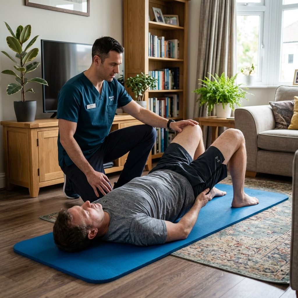
The exact mechanisms by which a spinal structural lesion causes radiating sciatic pain are deeply multifactorial, involving a complex interplay of both mechanical and profound biochemical components. Pure mechanical compression occurs when physical structures intrude into the critical, limited spaces of the spinal canal or intervertebral foramina. These structures most commonly include the displaced, herniated gelatinous material of a compromised intervertebral disc, but can also involve hypertrophic (enlarged) facet joints due to osteoarthritis, or a thickened, fibrotic ligamentum flavum. This direct, unyielding physical pressure on the delicate nerve root can physically deform the nerve fibers, dramatically disrupt the axoplasmic flow (the vital transport of cellular materials and organelles within the nerve axon), and impair normal electrical impulse conduction. This mechanical failure translates clinically into 'negative' neurological symptoms, such as profound numbness, loss of reflexes, and measurable motor weakness (paresis) in the muscles supplied by that specific nerve root.
However, it is widely recognized in modern spinal pathology that mechanical compression alone does not fully or adequately explain the intense, severe, and highly irritable pain experienced by many acute sciatica patients. The crucial concept of 'chemical radiculitis' is indispensable for understanding the acute phase of sciatic pain. The nucleus pulposus—the highly hydrated, gel-like center of the intervertebral disc—is avascular and isolated from the body's systemic immune system from birth. Consequently, it contains abnormally high levels of potent inflammatory mediators, most notably tumor necrosis factor-alpha (TNF-alpha), various interleukins, and phospholipase A2. When a significant annular tear allows this sequestered nucleus material to extrude and come into direct physical contact with the highly vascularized and sensitive spinal nerve root, the immune system recognizes it as a foreign invader. This triggers a massive, severe local inflammatory cascade and an autoimmune-like reaction, generating excruciating pain even in the complete absence of massive, space-occupying physical compression.
This overwhelming inflammatory response inevitably leads to significant intraneural edema (swelling) within the nerve root itself. Because the nerve root is confined within the already tight, bony tunnel of the intervertebral foramen, this internal swelling exponentially increases the pressure within the nerve, creating a vicious cycle of compression and inflammation. The inflamed nerve becomes pathologically hypersensitive, entering a state known as 'peripheral sensitization,' where even entirely normal, benign physiological movements or minor mechanical pressure variations can elicit severe, unremitting, shooting pain down the leg. If left untreated or mismanaged, chronic, low-grade compression and persistent inflammation can eventually stimulate fibroblasts to lay down scar tissue, leading to intraneural fibrosis. This permanent scarring physically alters the nerve's intrinsic biomechanical properties, destroying its vital elasticity and making it permanently less resilient and highly painful during the normal stretching required by daily activities, resulting in intractable, chronic sciatica.
Primary Etiologies and Modifiable Risk Factors for Sciatica
Lumbar disc herniation remains the single most prevalent and well-documented primary etiology for the development of classical sciatica, accounting for the vast majority of clinically significant radiculopathies. As individuals age, or secondary to repeated, incorrect biomechanical loading—such as heavy lifting with a flexed, twisted spine—the tough, multi-layered outer ring of the intervertebral disc (the annulus fibrosus) begins to weaken, degenerate, and develop micro-fissures. This structural compromise allows the highly pressurized, gel-like inner core (the nucleus pulposus) to bulge, protrude, or completely herniate outward. Due to the anatomical weakness of the posterior longitudinal ligament, this herniation typically occurs in a posterolateral direction—precisely the exact anatomical trajectory where the descending and exiting spinal nerve roots are located, leading to immediate, devastating mechanical compression and intense chemical inflammation. Additionally, the broader, natural aging process known as Degenerative Disc Disease (DDD) acts as a major precursor. As discs lose their essential hydration and structural height over decades, the complex biomechanics of the spinal segment are altered, leading to joint instability and the subsequent formation of reactive, compressive bone spurs (osteophytes) as the body attempts to artificially restabilize the failing joint.
Lumbar Spinal Stenosis represents another leading cause of severe sciatica, particularly prevalent within the geriatric population. This condition is characterized by a gradual, progressive narrowing of the central spinal canal or the lateral recesses and neural foramina. This insidious narrowing is typically the cumulative result of a degenerative cascade involving osteoarthritis of the spinal facet joints, progressive calcification and thickening of the spinal ligaments, and the bulging of collapsing, desiccated discs. Another significant structural etiology is 'spondylolisthesis,' a condition where one lumbar vertebra pathologically slips forward over the vertebra immediately below it, effectively guillotining the exiting nerve roots. This slippage can occur secondary to stress fractures in the pars interarticularis (isthmic spondylolisthesis, common in young athletes) or due to severe, advanced joint degeneration (degenerative spondylolisthesis, common in older adults). In all these structural pathologies, the end result is a critical loss of space for the neural elements.
A myriad of both modifiable and non-modifiable risk factors significantly exponentially increase an individual's susceptibility to developing debilitating sciatica. While age-related degenerative changes are ubiquitous and unavoidable, lifestyle factors play a profoundly deterministic role in spine health. Obesity and excessive body weight place immense, continuous, and unrelenting mechanical shearing and compressive forces on the lower lumbar discs, dramatically accelerating their structural breakdown and tripling the risk of herniation. Occupations demanding heavy physical labor, particularly those involving repetitive twisting of the trunk while carrying heavy, awkward loads, drastically elevate injury risk. Conversely, and equally damaging, are sedentary occupations that require prolonged, unbroken periods of sitting. Prolonged sitting chronically deactivates the critical core stabilizing musculature and subjects the lumbar discs to significantly higher sustained intradiscal pressure compared to standing, leading to accelerated disc failure. Furthermore, systemic factors such as smoking have been definitively proven to impair the already tenuous microvascular blood supply to the intervertebral discs, hindering their natural ability to heal from micro-traumas and accelerating the entire degenerative process.
Comprehensive Clinical Presentation: Radicular Pain to Paresthesia
The clinical presentation of true sciatica is remarkably distinct, diverse, and often so pathognomonic that a highly skilled, experienced clinician can accurately diagnose the specific, exact compressed nerve root based almost entirely on the patient's subjective description of their symptoms. The absolute hallmark symptom is unilateral 'radicular pain,' which is pain that precisely follows a specific, anatomically defined sensory pathway known as a dermatome. Patients characteristically describe this unique pain as sharp, searing, burning, or resembling a high-voltage electrical shock. It typically originates deep in the lower back or gluteal region, radiates forcefully downward through the posterior or lateral thigh, and frequently extends past the knee all the way into the calf, ankle, and toes. Crucially, the leg pain experienced in true sciatica is almost universally reported to be significantly more severe, agonizing, and disabling than any accompanying lower back pain.
Differentiating between an L5 and S1 radiculopathy is the absolute cornerstone of a precise clinical neurological assessment. When the L5 nerve root is structurally compressed, the radiating pain is heavily concentrated along the lateral (outer) aspect of the thigh and calf, sweeping across the dorsum (top) of the foot and terminating at the great toe. These patients frequently complain of profound numbness localized specifically in the web space between the first and second toes. Motor examination often reveals significant weakness in the extensor hallucis longus muscle, leading to difficulty or inability to extend the big toe or perform active dorsiflexion of the ankle, a condition clinically known as 'foot drop,' which causes them to drag their foot while walking. In stark contrast, an S1 nerve root compression produces pain that predominantly travels straight down the true posterior (back) of the thigh and the calf muscles, extending into the lateral border and sole of the foot. The classical neurological signs for an S1 lesion include a demonstrably diminished or completely absent Achilles tendon reflex, profound weakness in the powerful calf muscles (plantar flexors) leading to an inability to stand or walk safely on their tiptoes.
In addition to the hallmark radiating pain and motor weakness, sciatica patients persistently suffer from a distressing array of sensory disturbances broadly categorized as paresthesia. These manifest as relentless tingling, 'pins and needles' sensations, and areas of profound, 'dead' numbness. More complex neurological phenomena are also frequently observed, such as hyperalgesia (where a mildly painful stimulus is perceived as excruciatingly agonizing) and allodynia (where a completely non-painful stimulus, like the light brushing of clothing against the skin, elicits severe, sharp pain). Clinically, the constellation of sciatica symptoms is notoriously exacerbated by any physical maneuver that rapidly increases pressure within the abdominal cavity or the spinal canal itself (Valsalva maneuvers). Consequently, simple acts like coughing, sneezing, laughing, or bearing down during a bowel movement can trigger explosive, shooting pain down the leg. Furthermore, patients universally report significant pain exacerbation during prolonged sitting or when adopting a forward-flexed posture, while they may find substantial, temporary relief by lying flat in a supine position, which biomechanically unloads the distressed spinal segments.
Red Flag Symptoms Requiring Immediate Medical Attention
While the vast majority of sciatica and radiculopathy cases stem from benign, mechanical causes that respond exceptionally well to targeted, conservative physical therapy programs, clinicians must remain hyper-vigilant for a specific constellation of clinical signs and symptoms universally designated as 'Red Flags.' The presence of any of these red flags strongly indicates a high probability of a severe, potentially catastrophic, and limb- or life-threatening underlying medical emergency that absolutely mandates immediate, emergency surgical or specialized medical intervention to prevent irreversible, permanent neurological devastation. The most critical and feared of these absolute emergencies is 'Cauda Equina Syndrome,' a rare but devastating condition occurring when the entire bundle of nerve roots at the terminal end of the spinal cord (the cauda equina) is subjected to sudden, massive, and comprehensive compression, most frequently caused by a massive, central disc herniation.
The classic, definitive, and most critical symptoms of Cauda Equina Syndrome include a sudden, acute, or rapidly progressive loss of voluntary control over bowel or bladder functions. This autonomic failure may manifest clinically as severe urinary retention (an absolute inability to initiate a stream or empty the bladder), or alternatively, sudden urinary or fecal incontinence (an inability to hold urine or stool). Another paramount warning sign is the development of 'saddle anesthesia,' which is defined as a profound loss of sensation, or severe, dense numbness, distributed specifically in the anatomical areas that would rest on a horse's saddle—namely, the perineum, the genitals, the buttocks, and the medial aspects of the upper thighs. Furthermore, the rapid onset of bilateral motor weakness—meaning sudden, progressive, profound weakness affecting both legs simultaneously, making the patient acutely unable to bear weight, lift their feet, or walk—is an incredibly grave neurological sign demanding immediate emergency room evaluation and likely emergent decompressive surgery within hours.
Beyond the immediate structural threat of Cauda Equina Syndrome, both patients and healthcare providers must be acutely aware of other systemic red flags that could indicate a severe, non-mechanical etiology masquerading as simple sciatica. The presence of unrelenting, continuous, unremitting night pain that completely fails to improve with any position change or rest, especially when accompanied by unexplained, rapid, and significant weight loss, loss of appetite, or a known personal history of cancer, significantly raises the clinical suspicion for spinal malignancies, metastatic disease, or invasive tumors compressing the nerve roots. Similarly, the presence of an unexplained, persistent fever, profound chills, night sweats, or severe, localized spinal pain in a patient with a recent history of intravenous drug use, a recent invasive medical procedure, or a compromised, weakened immune system, strongly points toward the possibility of a highly destructive, deep spinal infection, such as bacterial discitis or a spinal epidural abscess. All of these severe, underlying systemic conditions must be definitively and urgently ruled out by a medical doctor through advanced diagnostics before any intensive, active physical therapy rehabilitation program can be safely initiated.
The Diagnostic Pathway: Clinical Evaluation to Advanced Imaging
The pathway to an accurate, definitive diagnosis of sciatica is a meticulous blend of clinical art and medical science, relying overwhelmingly on a highly detailed patient history and a comprehensive, systematic physical and neurological examination. The physical therapist's clinical evaluation is paramount in this phase. The assessment begins with an in-depth inquiry into the exact mechanism of injury, the specific chronicity and quality of the pain, and the precise mechanical factors that provoke or alleviate the symptoms. The core of the physical examination involves a battery of highly specific neurological provocative tests, meticulously designed to selectively place mechanical tension on the sciatic nerve to reproduce the patient's familiar symptoms, thereby confirming nerve root involvement. The most universally recognized and validated of these is the Straight Leg Raise (SLR) test. During the SLR, the clinician passively elevates the patient's straightened leg while they lie supine. If this maneuver elicits shooting, radicular pain traveling down the leg (not just tightness in the hamstrings) at an angle of elevation strictly between 30 and 70 degrees, it is considered a highly positive clinical indicator of lower lumbar (L5 or S1) nerve root compression.
To maximize diagnostic accuracy and rule out false positives, the clinician will invariably employ confirmatory provocative maneuvers. If the SLR is positive, the clinician may lower the leg slightly until the pain stops and then sharply dorsiflex the foot (Braggard's test) or apply direct pressure to the tibial nerve in the popliteal fossa (Bowstring sign); a return of the shooting pain definitively confirms neural tension. To evaluate the higher lumbar nerve roots (L3-L4), which form the femoral nerve, the clinician will utilize the Femoral Nerve Stretch Test, performed with the patient lying prone. A meticulous, thorough neurological examination is then conducted, which is essential for mapping the exact extent of nerve damage. This includes rigorous manual muscle testing of specific 'myotomes' to detect even subtle motor weakness, meticulous sensory testing of specific 'dermatomes' using light touch and pinprick to identify distinct areas of numbness, and the rigorous testing of deep tendon reflexes (such as the patellar and Achilles reflexes), where a diminished or absent reflex indicates significant, objective impairment in the neural conduction pathway.
While a masterful clinical examination is frequently sufficient to establish a highly reliable working diagnosis and safely initiate a targeted, evidence-based physical therapy program, advanced medical imaging technologies play an absolutely indispensable role in visualizing the actual structural pathology, confirming the clinical diagnosis, and planning more invasive medical or surgical interventions if conservative care fails. Magnetic Resonance Imaging (MRI) stands unchallenged as the absolute gold standard imaging modality for the comprehensive evaluation of the spine and all its intricate soft tissue structures. MRI provides incredibly detailed, high-resolution, three-dimensional visualization of the intervertebral discs, the exiting spinal nerve roots, the spinal cord itself, and the surrounding ligaments, making it the supreme tool for definitively identifying disc herniations, annular tears, and spinal stenosis. Computed Tomography (CT) scans may be utilized, particularly when evaluating complex bony abnormalities, fractures, or significant osteophyte formation. In highly complex, ambiguous cases where the clinical presentation is confusing, or where it is difficult to definitively distinguish between a spinal radiculopathy (nerve root issue) and a peripheral neuropathy (damage to the nerve further down the leg, such as in diabetes), the physician will order an Electromyography (EMG) combined with Nerve Conduction Studies (NCS). These functional, electrical tests precisely measure the speed and integrity of electrical signals traveling through the nerves and muscles, definitively confirming the exact location and physiological severity of the neural damage.
The Crucial Role of Conservative Management as First-Line Defense
The established medical paradigm for managing acute sciatica has undergone a massive, evidence-based revolution over the past few decades, completely abandoning the antiquated, deeply flawed historical practices that mandated extended periods of strict bed rest. Contemporary, rigorous clinical trials and medical consensus guidelines have unequivocally demonstrated that prolonged bed rest—defined as continuous rest exceeding 48 hours—provides absolutely no therapeutic benefit for sciatic pain. In stark reality, prolonged immobility is profoundly detrimental; it rapidly exacerbates spinal joint stiffness, accelerates the dangerous atrophy and profound weakening of the critical core stabilizing musculature, significantly increases the risk of deep vein thrombosis, and ultimately promotes the tragic transition from a manageable acute pain episode into a complex, devastating chronic pain syndrome. In direct opposition to bed rest, the modern, gold-standard approach champions 'Active Conservative Management' and intelligent activity modification as the absolute first and most effective line of defense against the debilitating symptoms of disc herniation and sciatic nerve compression.
The central, guiding philosophy of modern conservative management is encapsulated in the concept of 'Relative Rest.' This nuanced approach strongly encourages the patient to remain as physically active as their pain tolerance safely allows, continuing to perform basic, daily functional movements. However, it mandates the strict, disciplined avoidance of highly specific, biomechanically demanding activities that are known to heavily load the vulnerable spinal structures and violently exacerbate radicular symptoms—such as repetitive, loaded forward bending (spinal flexion), lifting heavy objects, twisting the spine under load, or enduring unbroken, prolonged periods of sitting. This highly structured activity modification is inextricably paired with a robust, empowering patient education program. A critical duty of the physical therapist is to thoroughly reassure the patient, helping them completely decouple the intense sensation of pain from the terrifying fear of ongoing, severe tissue damage. Patients must be educated that severe, shooting sciatic pain does not necessarily equate to a structurally unstable or 'broken' spine, and they must understand that the natural, unhindered biological course of a herniated disc is overwhelmingly favorable, leaning heavily toward natural healing and symptom resolution over time.
One of the most biologically fascinating, scientifically validated, and profoundly reassuring physiological facts that we extensively educate our patients about is the phenomenon of 'Spontaneous Disc Resorption.' When a portion of the nucleus pulposus herniates and extrudes beyond the protective annulus and into the spinal canal, the body's highly intelligent immune system immediately recognizes this displaced gel-like material as a foreign entity. The immune system then mounts a highly orchestrated, targeted response, deploying specialized cells called macrophages to aggressively attack, break down, and phagocytize the herniated disc fragment. Through this remarkable, natural biological process, the physical size of the herniated disc gradually but definitively shrinks and recedes over weeks and months, naturally and safely lifting the crushing mechanical pressure off the distressed nerve root. A comprehensive, expertly guided physical therapy program provides the absolute optimal, frictionless biomechanical environment to support, accelerate, and maximize this natural physiological healing process. By expertly managing the debilitating symptoms during this crucial waiting period, physical therapy successfully guides the vast majority of patients away from the significant risks and prolonged recoveries associated with unnecessary spinal surgeries.
Evidence-Based Physical Therapy: Core Principles in Decompression
Evidence-based physical therapy stands unequivocally as the primary, most vital, and most consistently successful pillar for achieving comprehensive, sustainable recovery from acute episodes of sciatica and lumbar disc herniations. The role of a highly trained, specialized physical therapist extends far beyond merely providing temporary, superficial pain relief modalities. Instead, the expert therapist conducts a profound, highly analytical biomechanical and functional assessment to identify, isolate, and systematically correct the deep-seated movement dysfunctions, muscular imbalances, and faulty kinematic patterns that originally led to the devastating structural failure of the disc. This rigorous rehabilitation program begins with a highly meticulous evaluation of the patient's specific movement impairments, identifying critical deficits in muscular strength and endurance, assessing joint mobility restrictions, and analyzing faulty neuromuscular control, ensuring that the resulting treatment plan is highly customized, aggressively targeted, and perfectly tailored to the unique mechanical presentation of the individual patient.
During the most acute, initial phases of sciatica, when the inflammatory chemical radiculitis is raging and the pain is agonizing and utterly overwhelming, the therapist may strategically employ specific, targeted passive physical modalities. The sole purpose of these modalities is to temporarily suppress the symptoms, quiet the highly irritable nervous system, and create a brief, precious 'window of opportunity' that allows the patient to tolerate the essential active movement exercises. This may involve the precise application of cryotherapy (ice) to powerfully constrict local blood vessels and reduce the debilitating inflammatory edema around the nerve root, or the use of Transcutaneous Electrical Nerve Stimulation (TENS) to bombard the sensory pathways and effectively block the transmission of severe pain signals to the brain. Furthermore, the therapist may utilize highly skilled, gentle manual therapy techniques, including low-grade, highly specific spinal joint mobilizations. These expert, hands-on techniques are designed not to forcefully realign bones, but to profoundly inhibit the massive, painful, and reactive muscle spasms that tightly guard the injured spine, thereby reducing the compressive forces acting on the damaged intervertebral discs.
However, as soon as the initial, acute wave of searing pain begins to subside, the entire focus of the rehabilitation program shifts decisively and aggressively toward active, patient-driven, movement-based interventions. The primary, overriding physiological and mechanical goal in this crucial phase is 'Unloading'—systematically removing the immense, pathological mechanical stress from the highly sensitized, compressed neural structures. This is expertly achieved through a carefully orchestrated, systematic progression of targeted stretching exercises aimed at restoring normal resting length to pathologically tight, restrictive muscle groups, such as the powerful hip flexors, the massive hamstring complex, and the deep, often-spasming piriformis muscle. Concurrently, these mobility exercises are seamlessly integrated with the early, highly precise initiation of neuromuscular control training. This specialized training is designed to re-educate the nervous system, ensuring that the critical stabilizing muscles surrounding the lumbar spine and pelvis fire in perfect, harmonious synchrony, thereby protecting the vulnerable spinal joints from aberrant, destructive movements and entirely eliminating the devastating micro-shearing forces that perpetuate disc injury and nerve irritation.
The McKenzie Method (MDT) and the Phenomenon of Directional Preference
The McKenzie Method, formally recognized worldwide as Mechanical Diagnosis and Therapy (MDT), stands as one of the most scientifically validated, universally respected, and clinically powerful assessment and treatment frameworks available for the management of spinal pain, radiating radiculopathy, and sciatica caused by intervertebral disc derangements. This unique, highly logical methodology relies entirely on observing the patient's immediate, objective, and reproducible symptomatic and mechanical responses to specific, repeated spinal movements or sustained postural positions. Unlike many passive treatment approaches, the MDT assessment categorizes the patient's clinical presentation into specific mechanical syndromes based strictly on how their pain behaves under dynamic loading. This rigorous diagnostic process reliably determines whether the severe sciatic symptoms are positively or negatively influenced by precise mechanical forces, thereby providing the clinician and the patient with a crystal-clear, measurable, and highly predictable roadmap for intervention, completely eliminating the dangerous guesswork often associated with spinal care.
The absolute core, defining principle, and most 'magical' clinical phenomenon utilized within the McKenzie Method is the discovery of 'Centralization' and the identification of the patient's 'Directional Preference.' Centralization is a thoroughly documented, highly reliable clinical phenomenon where the patient's severe, radiating pain—which may extend far down the leg, into the calf, or even to the toes—rapidly and progressively retreats up the leg, moving closer to the midline of the lower back, in direct response to a specific, repeated movement. The occurrence of this rapid centralization phenomenon during the initial assessment is considered an overwhelmingly positive prognostic indicator, signifying that the disc derangement is highly reducible and that conservative therapy will be exceptionally successful. The 'Directional Preference' is the precise, specific direction of movement (e.g., spinal extension or spinal flexion) that actively drives this centralization process and immediately, significantly reduces the intensity of the patient's radicular symptoms.
In the vast majority of classic lumbar disc herniations resulting in sciatica (specifically, posterior or posterolateral herniations), patients exhibit a powerful, undeniable directional preference for 'Extension' (bending backwards). These patients are meticulously instructed and trained to perform highly specific, repeated extension exercises, such as the prone press-up (repeatedly pushing the upper body up while keeping the pelvis flat on the floor). Biomechanically, these targeted extension forces compress the posterior aspect of the disc, theoretically actively driving the displaced, herniated nuclear gel anteriorly, away from the highly sensitive, compressed nerve roots, thereby effectively abolishing the mechanical pressure and reducing the inflammatory chemical radiculitis. Alongside extension protocols, patients are also taught how to actively self-correct a 'Lateral Shift'—a very common, painful, and involuntary postural deformity where the patient's torso is visibly shifted to one side in a desperate, subconscious attempt to mechanically decompress the trapped nerve. The ultimate power and brilliance of the McKenzie Method lie in its profound ability to empower the patient; by transforming them into an independent self-treater, they learn exactly how to use their body's own mechanics to rapidly abolish their pain, manage their symptoms independently, and prevent future recurrences without relying constantly on a healthcare provider.
Neural Mobilization: Advanced Nerve Gliding and Flossing Techniques
Neurodynamics, commonly referred to in clinical practice as Neural Mobilization or nerve flossing, represents an advanced, highly specialized, and absolutely critical set of therapeutic techniques in the comprehensive rehabilitation of sciatica. These interventions are founded on the fundamental physiological and biomechanical understanding that the entire human nervous system is a continuous, unbroken tissue tract. To function normally and painlessly, this vast neural network must possess the essential ability to slide, glide, and elongate smoothly and freely through its complex surrounding anatomical interfaces—such as muscles, fascia, and bony tunnels—during every single movement we make. When a spinal nerve root suffers severe inflammation and physical compression due to a herniated disc, the body's healing response inevitably forms microscopic adhesions and restrictive scar tissue around the nerve, severely binding it down and destroying its normal, vital mobility. This pathological restriction causes the nerve to undergo massive, abnormal mechanical tension during simple, daily movements, generating explosive, searing pain signals and severely retarding the overall physiological recovery process.
To aggressively combat this neural tethering and actively restore healthy, pain-free nerve function, physical therapists expertly employ two primary, highly specific categories of neurodynamic techniques: 'Nerve Sliders' (or Nerve Flossing) and 'Nerve Tensioners.' Nerve Sliders are specifically designed to maximize the total excursion and sliding movement of the entire sciatic nerve tract through its anatomical pathway, while intentionally keeping the overall mechanical tension on the delicate nerve fibers to an absolute minimum. This ingenious, gentle effect is achieved by moving two joints simultaneously in a highly coordinated fashion; the therapist (or the patient, once trained) applies tension to one end of the nerve tract by moving a joint, while simultaneously releasing tension at the other end by moving an opposing joint in a specific direction. This coordinated 'push-pull' mechanism ensures that the nerve is smoothly 'flossed' back and forth within its protective sheath, safely and effectively breaking up constrictive adhesions, improving intraneural blood flow, and powerfully flushing out stagnant, inflammatory chemicals without ever provoking or worsening the patient's severe pain.
Once the acute, highly irritable phase of sciatica has completely subsided, and the nerve has regained a substantial degree of pain-free sliding mobility, the therapist will carefully, systematically, and highly cautiously progress the patient to 'Nerve Tensioners.' In stark contrast to sliders, tensioners are explicitly designed to apply a gradual, measured, and deliberate mechanical stretch to the nerve tissue itself. The physiological goal of this advanced technique is to actively restore the nerve's intrinsic, microscopic elasticity (neuro-elasticity), ensuring it can fully tolerate the extreme mechanical elongation required during high-level athletic activities and vigorous daily movements without sustaining micro-trauma or generating pain. It is an absolute, critical clinical imperative that all neurodynamic techniques be applied with exceptional caution and masterful clinical skill. Applying excessive tension, or utilizing aggressive nerve stretching prematurely during the acutely inflamed phase of chemical radiculitis, will inevitably severely aggravate the already hyper-sensitive nerve, triggering a massive flare-up of excruciating symptoms. These powerful techniques are eventually seamlessly integrated into the patient's independent home exercise routine, ensuring the continuous, lifelong maintenance of optimal neural mobility and preventing the devastating return of chronic stiffness and nerve pain.
Postural Re-education and Ergonomic Optimization for Daily Living
Achieving a complete, sustainable, and lifelong recovery from severe sciatica is virtually impossible without aggressively addressing, correcting, and permanently modifying the deeply ingrained, faulty daily postural habits and flawed biomechanics that place relentless, pathological mechanical stress on the vulnerable lumbar intervertebral discs. Prolonged sitting, particularly in environments that fail to provide adequate, anatomically correct support, is widely recognized as one of the most highly destructive, insidious factors contributing to catastrophic spinal failure. Highly precise, in-vivo biomechanical measurements have definitively proven that the internal pressure within the lumbar intervertebral discs (intradiscal pressure) skyrockets dramatically during sitting, particularly when an individual adopts a 'slouched,' forward-flexed posture, compared to the pressure experienced while standing upright or lying supine. This massive, sustained, and highly abnormal elevation in internal disc pressure forcefully drives the gelatinous nucleus pulposus backwards, constantly stressing the weakened posterior annulus, directly exacerbating existing disc herniations, and drastically intensifying the mechanical compression on the desperate, trapped sciatic nerve roots.
A massive, foundational component of the comprehensive physical therapy plan is dedicated to the thorough, exhaustive education and practical training of the patient in the critical principles of ergonomics and postural optimization. Patients are rigorously trained to understand the absolute necessity of maintaining the spine's natural, shock-absorbing curves—most importantly, the 'Lumbar Lordosis' (the natural inward curve of the lower back)—at all times, regardless of the activity. This comprehensive guidance involves detailed, personalized instruction on how to correctly setup an ergonomic workstation, ensuring optimal chair height, proper monitor placement, and the absolutely non-negotiable use of firm lumbar supports (lumbar rolls) to perfectly fill the void between the lower spine and the chair, physically preventing the pelvis from rotating backwards into a highly destructive flexed position. Furthermore, patients are strongly conditioned to adopt a dynamic strategy of 'Postural Variability.' They are taught that no single posture is healthy if maintained indefinitely, and are instructed to rigorously avoid remaining in any static position for longer than 30 to 40 minutes, integrating frequent, mandatory micro-breaks to stand, stretch, and walk, thereby dramatically reducing the cumulative, crushing mechanical fatigue on the spinal tissues.
In addition to completely overhauling their sitting habits, intense, repetitive focus is placed on completely retraining the patient's biomechanics for lifting and carrying objects, regardless of weight. Patients are systematically taught, drilled, and trained to flawlessly execute the 'Hip Hinge' mechanic. This essential movement pattern ensures that the massive, immensely powerful muscles of the glutes and hamstrings act as the primary engines for lifting, completely sparing the highly vulnerable, easily damaged lumbar spine from dangerous flexion under load. Patients learn the critical, uncompromising rule of keeping all lifted objects as close to their body's center of mass as physically possible. This dramatically reduces the biomechanical 'lever arm,' thereby minimizing the massive, destructive rotational torque and shearing forces that are highly capable of tearing spinal ligaments and violently rupturing intervertebral discs. By deeply internalizing and rigorously applying these ergonomic principles, patients successfully transform their daily environment and habits from primary sources of catastrophic spinal injury into highly supportive, protective foundations for sustainable recovery and lifelong prevention.
Core Strengthening and Stabilization: Building a Resilient Spinal Support System
The modern, highly advanced, and scientifically validated concept of 'Core Stabilization' and spinal strengthening completely discards and transcends the antiquated, highly destructive traditional approaches that solely focused on superficial abdominal exercises, such as crunches and sit-ups. Extensive biomechanical research has proven that these outdated exercises actually generate massive, dangerous compressive forces on the intervertebral discs, actively worsening herniations and completely failing to provide the specific, necessary stability the injured spine desperately requires. Instead, cutting-edge rehabilitation programs focus entirely on the precise reactivation, neuro-muscular re-education, and endurance training of the spine's 'Deep Stabilizing System.' This critical, intricate muscular corset consists primarily of the Transversus Abdominis (acting as a powerful, deep, circumferential belt), the Lumbar Multifidus (providing microscopic, segment-by-segment control connecting each individual vertebra), the robust Pelvic Floor muscles from below, and the crucial Respiratory Diaphragm from above. Together, these deep, hidden muscles function as a brilliant, internal anatomical brace, working in perfect concert to provide dynamic stability and absolute micro-control to each individual segment of the spine.
The primary physiological and mechanical goal of training this deep stabilizing system is absolutely not to build massive, bulky muscles, but rather to flawlessly restore 'Motor Control'—the nervous system's ability to activate the right muscles, at the exact right time, with the precise amount of force. The vast majority of patients suffering from chronic sciatica and disc pathologies exhibit a highly documented, profound delay in the automatic, reflex-driven activation of these deep muscles (the feed-forward mechanism) prior to initiating any movement of the arms or legs. This critical neurological delay leaves the spinal joints completely unprotected and highly vulnerable to devastating micro-shearing forces and micro-traumas, which relentlessly perpetuate the disc injury and nerve irritation. Through highly specialized, low-level exercises and synchronized diaphragmatic breathing techniques, physical therapists literally 'reprogram' the patient's central nervous system to instinctively and automatically fire this deep internal corset a fraction of a second before moving their limbs. This guarantees the optimal regulation of 'Intra-abdominal Pressure,' creating a powerful, protective pneumatic cylinder of support that dramatically unloads the spine and shields it from high-velocity forces.
Once the patient has successfully mastered the flawless, pain-free, automatic activation of this deep, foundational stabilizing system, the strengthening program is systematically, carefully, and aggressively progressed to incorporate the larger, superficial 'global' muscles responsible for producing powerful movements and bearing heavy loads—such as the rectus abdominis, the massive gluteal complex, and the powerful erector spinae group. These advanced, high-level exercises progressively integrate highly dynamic, complex limb movements while the patient must simultaneously maintain absolute, rigid stability of the lumbar core, perfectly simulating the intense, unpredictable mechanical demands of heavy daily activities, demanding occupations, and high-level athletic pursuits. Ultimately, building a profoundly strong, functionally stable, and neurologically perfectly-timed core provides an impenetrable, long-lasting protective shield for the entire spine, dramatically and permanently reducing the probability of the intervertebral discs and delicate nerve roots ever succumbing to a devastating future relapse.
Medical Management and Pharmacological Interventions Complementing Therapy
In severe, acute clinical scenarios where sciatica presents with agonizing, overwhelming, and unendurable pain that completely incapacitates the patient and utterly precludes their ability to actively participate in essential physical therapy and rehabilitation, specialized medical interventions and targeted pharmacological management play an absolutely vital, complementary role. It is imperative to understand that the primary goal of pharmacological management is not to cure the underlying mechanical structural fault or heal the herniated disc—that is the exclusive domain of time and mechanical rehabilitation—but rather to aggressively suppress the overwhelming pain response and significantly blunt the massive, acute inflammatory storm. This strategic suppression aims to reduce symptoms to a tolerable level, creating a crucial therapeutic window that allows physical interventions to be effectively applied. The first line of pharmacological defense typically involves the strategic, scheduled use of Non-Steroidal Anti-Inflammatory Drugs (NSAIDs) at prescription doses to powerfully inhibit the systemic inflammatory cascade and reduce the local edematous swelling heavily compressing the nerve root. Additionally, short, highly controlled courses of muscle relaxants may be prescribed to aggressively combat the severe, agonizing, and highly reactive reflex muscle spasms that frequently and painfully lock the injured spine during acute sciatic episodes.
For patients suffering from severe, intractable radicular pain and profound neuropathy—often described as a relentless burning, searing, or electrical shock sensation that stubbornly fails to respond to traditional, standard analgesics—specialized physicians may strategically prescribe advanced Neuropathic Pain Modulators, such as Gabapentin or Pregabalin. These powerful, highly specific medications act directly on the central and peripheral nervous systems, effectively altering nerve conduction and significantly calming the hyper-excitable, rapid-firing electrical signals generated by the severely irritated, compressed nerve root. In documented cases of massive, acute, and severely debilitating chemical radiculitis, physicians may cautiously consider prescribing a short, aggressive, tapering dose of oral corticosteroids (such as a Medrol dose pack). The objective here is the rapid, systemic, and overwhelming suppression of the local, destructive immune response and severe inflammation, although this aggressive approach is used highly selectively and judiciously due to the significant risk of broad, undesirable systemic side effects associated with high-dose steroids.
When all conservative oral medications comprehensively fail to provide adequate, necessary relief, and the patient's agonizing pain continues to severely block any meaningful rehabilitation progress, an Epidural Steroid Injection (ESI) emerges as a highly effective, targeted interventional medical procedure. This precise procedure, invariably performed under the exacting guidance of live fluoroscopic X-ray to ensure absolute millimeter accuracy, involves the direct injection of a highly concentrated, potent dose of long-acting anti-inflammatory corticosteroids, often combined with a fast-acting local anesthetic, directly into the epidural space immediately surrounding the inflamed, trapped, and desperate nerve root. It is crucial to educate the patient that an ESI is not a permanent 'cure' for the structural herniation itself; rather, it is designed to provide a profound, substantial, yet temporary 'window of opportunity' characterized by dramatic pain relief. During this highly valuable, golden, pain-free window, the physical therapist and the patient must aggressively and intensively capitalize on the relief to rapidly advance through the complex biomechanical rehabilitation program, correct the underlying postural faults, and restore essential tissue flexibility, thereby ensuring that when the injection's effects eventually wear off, the mechanical cause of the pain has been fundamentally resolved.
Surgical Considerations for Refractory Sciatica and Minimally Invasive Procedures
Despite the vast, overwhelming success and powerful capabilities of comprehensive, evidence-based conservative physical therapy—which has been definitively proven to successfully manage and resolve over 90% of sciatica and lumbar disc herniation cases over time—a small, distinct percentage of patients with highly specific, complex, and unyielding clinical presentations will ultimately require meticulous evaluation and definitive surgical intervention. The critical, clinical indications that strongly drive a spinal surgeon to recommend operative intervention are based on extremely rigorous, objective medical and neurological criteria. These strict indications include the absolute, comprehensive failure of exhaustive, high-quality conservative care over a minimum period of 6 to 12 weeks to achieve any meaningful, tolerable reduction in severe, incapacitating pain, significantly diminishing the patient's quality of life. Furthermore, surgery is strongly indicated upon the alarming emergence of a rapidly progressive, deteriorating neurological deficit—manifesting clinically as a swift, measurable, and significant increase in leg weakness or profound foot drop. Most critically, any presentation of the aforementioned emergency 'Red Flags,' particularly the onset of Cauda Equina Syndrome with bowel or bladder dysfunction, dictates immediate, emergency, life-altering surgical decompression within hours to prevent permanent paralysis.
For the vast majority of surgical candidates suffering from classical sciatica caused specifically by a localized lumbar disc herniation, a 'Microdiscectomy' represents the absolute, gold-standard surgical procedure. This highly refined, elegant operation is characterized by its exceptionally minimally invasive nature. The specialized neurosurgeon or orthopedic spine surgeon utilizes an extremely small, targeted incision and employs sequential, tubular muscle dilators to gently separate, rather than aggressively cut, the thick spinal musculature. Utilizing a powerful, high-definition operating microscope for supreme visualization, the surgeon meticulously accesses the spinal canal and delicately, precisely removes only the extruded, herniated fragment of the disc material that is actively, mechanically crushing the spinal nerve root. Crucially, the surgeon exercises extreme care to leave the vast majority of the healthy, structurally sound inner disc and the tough outer annulus entirely intact. This vital preservation ensures that the disc can continue to perform its essential, lifelong biomechanical functions of bearing immense spinal loads and effectively absorbing high-impact shocks.
In more complex, challenging clinical scenarios where the patient's severe sciatica is the direct result of advanced, widespread Lumbar Spinal Stenosis—characterized by massive bone spurs and thickened ligaments—rather than a simple, isolated soft-tissue disc herniation, a significantly more extensive, decompressive surgical procedure known as a 'Laminectomy' may be absolutely required. This comprehensive operation involves the careful, calculated surgical removal of portions of the vertebral bone (the lamina) and the aggressive excision of overgrown, hypertrophied spinal ligaments to significantly widen the spinal canal and permanently provide ample, unrestricted space for the compressed neural elements. It is of the utmost, critical importance to emphatically stress to every single patient a fundamental medical truth: spinal surgery, even when flawlessly executed, is at its core merely a mechanical, structural decompression to relieve immediate pressure. To achieve a truly spectacular, long-term functional recovery and to confidently, safely return to a highly active, demanding lifestyle, comprehensive, rigorous post-operative physical therapy and extensive rehabilitation remain absolutely, non-negotiably indispensable to restore lost muscle strength, rebuild joint mobility, and permanently correct the faulty movement patterns that contributed to the initial spinal failure.
Comprehensive Prevention Strategies and Long-term Management
The successful, long-term management of sciatica and the permanent prevention of recurring lumbar disc herniations is not a passive event, but rather an ongoing, highly active process that demands a resolute, lifelong commitment to comprehensive lifestyle modification and the unwavering adoption of health behaviors deeply rooted in sound biomechanical principles. Maintaining a healthy, optimal body weight stands as an absolute, paramount priority in any effective, long-term spinal prevention strategy. Every single additional kilogram of excess body weight, particularly adipose tissue concentrated heavily in the central abdominal region, translates biomechanically into massive, continuous, and highly destructive compressive and shearing forces acting directly on the vulnerable lower lumbar intervertebral discs. This relentless, excessive mechanical loading dramatically accelerates the natural degenerative wear-and-tear process and exponentially increases the probability of suffering a catastrophic, future disc herniation.
Furthermore, meticulous attention to proper nutrition and aggressive, consistent systemic hydration are critical, yet frequently overlooked, foundational elements essential for maintaining optimal spinal health and preserving the structural integrity of the intervertebral discs. It is a vital physiological fact that mature, adult intervertebral discs possess absolutely no direct blood supply of their own. Instead, they rely completely and exclusively on a complex, motion-dependent physiological process known as 'imbibition' to absorb essential nutrients, oxygen, and vital water from the surrounding tissues, and to expel toxic metabolic waste products. This delicate process is heavily dependent on regular, dynamic spinal movement and the presence of abundant systemic hydration. Chronic, systemic dehydration directly and rapidly leads to the progressive desiccation (drying out) and dangerous shrinking of the discs. This loss of vital water content severely compromises the disc's elasticity, destroys its critical shock-absorbing capacity, and renders the tough outer annulus incredibly brittle, fragile, and highly susceptible to catastrophic tearing under even the most minor, trivial mechanical stresses.
Finally, aggressively addressing and successfully conquering the profound psychological and cognitive barriers associated with chronic pain management—most specifically, overcoming the debilitating, paralyzing fear of movement known clinically as 'Kinesiophobia'—is an absolutely integral, non-negotiable component of a truly comprehensive, holistic spinal rehabilitation program. Countless patients, deeply traumatized by the memory of excruciating, searing sciatic pain, unconsciously develop a severe, pathological fear of re-injury. Consequently, they compulsively avoid physical activity and severely restrict their normal movements. This fear-avoidance behavior initiates a tragic, highly destructive downward spiral resulting in rapid, severe muscle atrophy, profound joint stiffness, and a paradoxically massive increase in overall pain sensitivity and functional disability. Through expertly guided, cognitively-informed physical therapy, integrating principles of Cognitive Behavioral Therapy (CBT), patients are scientifically, safely, and gradually re-introduced to normal, graded physical activity. They are empowered to confidently perform healthy, dynamic spinal movements without fear, successfully shifting their mindset from that of a fragile, dependent patient to a resilient, active individual who confidently and independently manages their spine health, representing the ultimate, triumphant goal of our comprehensive rehabilitation program.
Unified Home Physical Therapy Standards & Geographical Coverage
Bidayah Center delivers highly structured home rehabilitation and clinical conditioning across all served territories, including Jeddah, Makkah, and Qatif districts. We deploy licensed physical therapists directly to your home, ensuring that you receive the same standards of care, specialized portable modalities, and treatment protocols as premier inpatient facilities.
Our medical coordinators screen each referral and coordinate field operations using regional dispatch networks to ensure prompt scheduling and strict adherence to appointment times. We maintain active communication channels with orthopedic surgeons and neurologists at major local hospitals to coordinate care.
While this generic page outlines the core clinical details of this service, we invite you to choose your specific city page. Doing so allows you to explore local neighborhood guidelines, read region-specific FAQs, and coordinate with male or female physical therapists according to the clinical needs of your family.