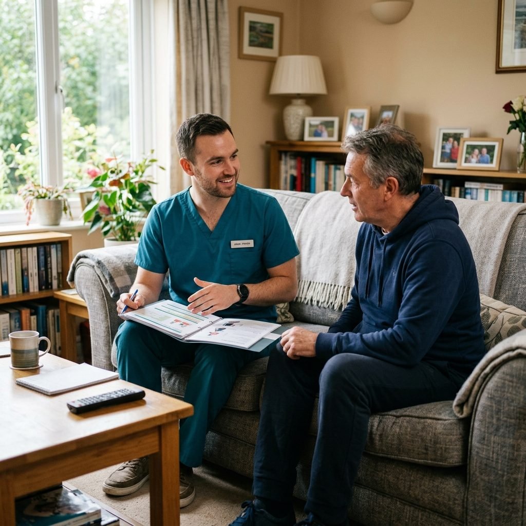
A comprehensive home safety and accessibility assessment is a cornerstone of preventive geriatric and rehabilitative medicine. Clinical research consistently demonstrates that structured environmental modifications reduce accidental falls by up to 40% in vulnerable populations. Our physical therapists evaluate the dynamic interaction between the patient's biomechanical capacity and the architectural features of their living space, delivering detailed blueprints for a safer home.
1. Preventive Clinical Philosophy of Home Safety Assessment
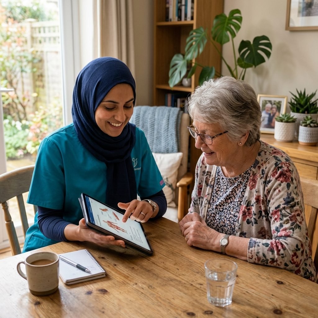
The clinical philosophy behind home accessibility assessments recognizes that modifying the physical environment is just as vital for rehabilitation as treating the patient's internal physical impairments. Adaptive spaces compensate for age-related sensory and motor declines.
Our objective is to achieve a harmonious alignment between the patient's physical limits and their home's spatial design. This alignment minimizes pathological loading on joints, reduces physical exhaustion, and allows patients to navigate their homes without fear of injury.
Mitigating environmental hazards directly reduces anxiety and fear of falling for both patients and their families. This psychological security encourages older adults to participate in daily activities, preventing the cascade of physical deconditioning and social isolation.
Within our integrated rehabilitation model, we view the home as an active therapeutic tool. A modified home environment supports the consistent execution of prescribed exercises and daily functional tasks, accelerating recovery and ensuring long-term safety.
2. Clinical-Environmental Assessment Protocol (STEADI Framework)
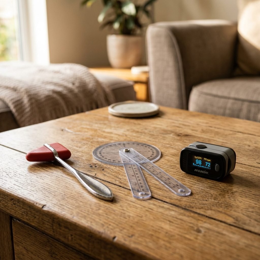
We utilize the medically endorsed Stopping Elderly Accidents, Deaths, and Injuries (STEADI) framework developed by the CDC. This comprehensive system combines physical screening of the patient with a detailed architectural check of their living spaces.
The physical screening includes testing the patient's visual acuity and contrast sensitivity, evaluating static and dynamic balance (such as the Berg Balance Scale), assessing orthostatic blood pressure changes, and taking a detailed history of any previous fall events.
The environmental inspection uses a clinical checklist to evaluate home entrances, stairwells, hallway widths, lighting systems, bedroom layouts, kitchen setups, and bathroom designs, identifying architectural barriers and potential trip hazards.
We analyze the biomechanical interaction between the patient and their furniture, ensuring that daily tasks, such as rising from a chair or reaching for items, can be performed safely without excessive spinal flexion or dangerous loss of balance.
3. Assessment and Modification of Entrances, Hallways, and Pathways
Entrances and hallways represent the primary transition zones where fall risks are elevated. We evaluate the stability of outdoor pathways, identify uneven steps or high thresholds, and recommend installing ramps with clinically safe slopes and handrails.
Within the home, we ensure that hallways are wide enough for assistive devices and free of obstacles like electrical cords, decorative furniture, or loose rugs. Unsecured rugs are a primary trigger for slips and trips in older adults.
We assess lighting levels in transitions and recommend installing motion-activated nightlights. Proper pathway lighting is essential for preventing disorientation and falls during nighttime bathroom visits, ensuring safety when ambient light is low.
We verify that sturdy handrails are installed on both sides of stairwells and along long corridors. We ensure the handrails are at an anatomically correct height (typically 34 to 38 inches) and offer a secure grip to support independent mobility.
4. Bedroom Layout Optimization and Functional Furniture Placement
The bedroom is a common site for fall injuries, particularly during nighttime transitions or hurried transfers. We measure the patient's bed height, ensuring that their feet rest flat on the floor when sitting at the edge of the mattress.
We recommend positioning the bed to allow a clear, unobstructed path to the bathroom. We eliminate all trip hazards along this route and ensure that essential items, such as glasses, medications, and phones, are within easy reach on a stable nightstand.
We optimize the heights of closets and drawers to prevent the patient from needing to bend down or reach high. We recommend firm mattresses that support spinal alignment and facilitate independent rolling and sitting up.
We ensure that bedside light controls are accessible from the bed, preventing the patient from moving in the dark. We also train patients on the proper placement of assistive devices next to the bed to ensure they are immediately available upon waking.
5. Bathroom Modifications and Advanced Slip-and-Fall Mitigation
Bathrooms present significant safety challenges due to moisture and slippery surfaces. Our assessment identifies precise locations for installing heavy-duty grab bars next to the toilet and inside the shower, ensuring they are anchored into wall studs.
We recommend using specialized shower chairs with slip-resistant rubber feet, combined with raised toilet seats to reduce knee and hip joint compression during transfers. Raised seats are critical for patients with joint degeneration or weakness.
We evaluate flooring surfaces and recommend installing non-slip rubber mats inside the tub and on bathroom floors. We advocate for handheld showerheads that allow the patient to bathe safely while seated, reducing fatigue and fall risks.
We ensure that shower thresholds are minimized or eliminated. Recommending zero-threshold, curbless walk-in showers is a primary strategy for preventing trips during entry and exit for patients with gait limitations.
6. Kitchen Accessibility and Ergonomic Appliance Integration
Kitchen tasks require functional reach, balance, and fine motor control. We evaluate the storage of frequently used items and recommend placing them in cabinets located between waist and shoulder height, eliminating the need to bend or climb.
We evaluate appliance safety and accessibility, recommending workspaces that allow the patient to prepare meals while safely seated. We emphasize the necessity of non-slip, easily cleanable flooring to manage accidental spills immediately.
For patients with arthritic joints or weak grip strength, we recommend ergonomic kitchen utensils with enlarged, textured handles. These design modifications reduce joint stress and support independence in meal preparation.
We review kitchen ventilation, emergency exits, and the accessibility of fire safety equipment. Ensuring that the patient can manage basic kitchen tasks safely and handle emergencies independently is vital for their safety and autonomy.
7. Lighting Systems, Contrast Sensitivity, and Visual Guidance
Lighting and visual contrast are critical for guiding movement and maintaining postural control. We measure lux levels throughout the home, identifying dark areas or harsh shadows that can distort spatial perception and lead to missteps.
We recommend applying high-contrast paint or tape to the edges of steps and thresholds to assist patients with low vision. Clear contrast around light switches and door handles also helps patients find them without unbalanced reaching.
We minimize glare from polished floors and unshaded windows, which can temporarily blind patients with cataracts or macular degeneration. We recommend anti-glare window coverings and positioning light sources to provide diffuse illumination.
Optimizing the home's visual environment helps patients judge depth, identify obstacles, and navigate level changes accurately. This support reduces motor errors and prevents collisions and falls caused by visual limitations.
8. Evaluation of Assistive Devices and Environmental Compatibility
Our assessment includes verifying that the patient's assistive devices (such as walkers, rollators, and wheelchairs) are compatible with the home's layout, checking doorway clearances and flooring transitions.
We inspect the rubber tips of canes and walkers for wear, replacing them to ensure optimal friction on smooth floors. We adjust the heights of these devices to match the patient's anatomical landmarks, promoting erect posture.
We recommend widening doorways that are narrower than the standard clearance required for wheelchairs (minimum of 32 inches). We also advocate for removing raised thresholds or replacing them with low-profile transitions.
We provide hands-on training for the patient and their family on using assistive devices safely at home. We teach them how to navigate tight spaces, perform turns, and transition between different flooring types without losing stability.
9. Environmental Modification Action Plan and Implementation Guide
Following the assessment, we deliver a detailed report detailing all identified risks, accompanied by practical recommendations for home modifications categorized by priority, clinical urgency, and estimated cost.
The action plan includes immediate, low-cost modifications (such as removing throw rugs, adding nightlights, and rearranging furniture) and structural modifications (such as installing grab bars, building ramps, and widening doors).
We provide a directory of qualified contractors specializing in accessibility modifications to ensure quality work and compliance with safety standards. Our therapists are available to consult during installation to verify accuracy.
This modification guide provides a sustainable path toward aging in place, reducing healthcare costs associated with fall injuries. Modifying the home is a proactive investment in the patient's long-term health and independence.
10. Behavioral Adaptation and Caregiver Safety Protocols
Successful home modification requires ongoing behavioral changes by the patient and their family. We complement physical changes with training on safe habits, helping patients integrate preventive practices into their daily routines.
We instruct patients to avoid high-risk behaviors, such as rushing to answer the phone, reaching for high shelves without a stable step stool, walking in dark hallways, or walking in socks without slip-resistant treads.
We train caregivers to maintain an organized home, keeping pathways clear of clutter and toys. We teach them to check for wet floors in kitchens and bathrooms and to ensure the patient wears a gait belt when needed.
Fostering these safe habits transforms home modifications into a comprehensive, family-wide safety culture. This behavioral support ensures that the physical changes to the home continue to protect the patient from injury over time.
11. Clinical Glossary of Physical Therapy and Rehabilitation Terms
This glossary provides essential medical and clinical definitions that form the basis of our home assessment protocols. Understanding these terms helps patients and caregivers comprehend the rehabilitation plan.
- ✓Humerus: The long bone of the upper arm, extending from the shoulder to the elbow and providing muscular support.
- ✓Radius: The lateral bone of the forearm, situated on the thumb side, facilitating pronation and supination.
- ✓Ulna: The medial bone of the forearm, extending parallel to the radius and forming the elbow joint.
- ✓Femur: The longest and strongest bone in the human body, connecting the pelvis to the knee and bearing weight.
- ✓Tibia: The larger medial bone of the lower leg, commonly called the shinbone, bearing most mechanical weight.
- ✓Fibula: The slender lateral bone of the lower leg, running parallel to the tibia and stabilizing the ankle.
- ✓Clavicle: A curved long bone connecting the sternum to the scapula, protecting superior vessels and nerves.
- ✓Scapula: A flat, triangular bone located on the posterior aspect of the shoulder girdle, assisting arm motion.
- ✓Sternum: A flat bone located in the middle of the anterior chest wall, anchoring the ribs for protection.
- ✓Ribs: Curved bones forming the thoracic cage, protecting internal organs and assisting in respiration.
- ✓Pelvis: A bony basin connecting the spine to the lower limbs and supporting abdominal visceral organs.
- ✓Sacrum: A triangular bone formed by fused sacral vertebrae, situated at the base of the vertebral column.
- ✓Coccyx: The small terminal portion of the spinal column, formed by fused rudimentary vertebrae.
- ✓Vertebra: An individual bone of the spinal column, enclosing and protecting the spinal cord.
- ✓Cervical Vertebrae: The first seven vertebrae of the spinal column, supporting the skull and head mobility.
- ✓Thoracic Vertebrae: The twelve middle vertebrae of the spine, articulating with the ribs to form the back.
- ✓Lumbar Vertebrae: The five lower vertebrae of the spine, bearing the greatest mechanical weight and load.
- ✓Cranium: The bony structure enclosing and protecting the brain and sensory organs, including facial bones.
- ✓Mandible: The only mobile bone of the skull, essential for mastication and speech production.
- ✓Maxilla: A fixed bone forming the upper jaw, nose, and the floor of the orbital cavities.
- ✓Zygomatic: The cheekbone forming the lateral portion of the eye orbit and facial structure.
- ✓Sphenoid: A complex bone at the skull base, articulating with and binding almost all other cranial bones.
- ✓Ethmoid: A light bone separating the nasal cavity from the brain and forming part of the orbit.
- ✓Temporal: A bone situated at the sides and base of the skull, housing the auditory organs.
- ✓Frontal: The bone forming the forehead, anterior cranial roof, and superior eye orbits.
- ✓Parietal: A paired bone forming the sides and roof of the cranium, protecting the cerebral cortex.
- ✓Occipital: A bone situated at the posterior-inferior region of the skull, containing the foramen magnum.
- ✓Carpals: Eight small bones forming the wrist joint, enabling multidirectional manual flexibility.
- ✓Metacarpals: Five bones forming the palm of the hand, connecting the wrist to the digits.
- ✓Phalanges: Small bones forming the digits of hands and feet, facilitating grip and prehension.
- ✓Tarsals: Seven bones forming the ankle and rearfoot, bearing the initial mechanical load of walking.
- ✓Metatarsals: Five long bones forming the arch of the foot, connecting the ankle to the toes.
- ✓Patella: A small sesamoid bone situated anterior to the knee joint, protecting it and enhancing quadriceps pull.
- ✓Biceps Brachii: A two-headed muscle on the anterior arm acting to flex the elbow and supinate the forearm.
- ✓Triceps Brachii: A three-headed muscle on the posterior arm acting to extend the elbow joint.
- ✓Deltoid: The triangular muscle covering the shoulder, acting to abduct, flex, and extend the arm.
- ✓Pectoralis Major: A large chest muscle acting to adduct, flex, and medially rotate the humerus.
- ✓Trapezius: A broad back muscle controlling scapular elevation, depression, retraction, and rotation.
- ✓Latissimus Dorsi: A broad back muscle acting to extend, adduct, and medially rotate the arm.
- ✓Rectus Abdominis: The straight abdominal muscle acting to flex the trunk and stabilize the pelvis.
- ✓Obliques: Lateral abdominal muscles facilitating trunk rotation, lateral flexion, and intra-abdominal pressure.
- ✓Gluteus Maximus: The largest gluteal muscle acting to extend the hip and maintain upright posture.
- ✓Sartorius: The longest muscle in the body, running across the thigh to flex the hip and knee.
- ✓Gracilis: A medial thigh muscle acting to adduct the hip and assist in knee flexion.
- ✓Iliopsoas: A primary hip flexor muscle composed of the psoas major and iliacus muscle units.
- ✓Piriformis: A deep gluteal muscle acting to laterally rotate the hip; can compress the sciatic nerve.
- ✓Supraspinatus: A rotator cuff muscle initiating arm abduction and stabilizing the humeral head.
- ✓Infraspinatus: A rotator cuff muscle acting to laterally rotate the arm and stabilize the glenohumeral joint.
- ✓Subscapularis: A rotator cuff muscle acting to medially rotate the arm and stabilize the shoulder joint.
- ✓Teres Minor: A small rotator cuff muscle assisting in external rotation and stability of the humerus.
- ✓Radial Nerve: A nerve innervating the extensor muscles of the arm, forearm, and hand.
- ✓Ulnar Nerve: A nerve running along the elbow, innervating the intrinsic hand and finger muscles.
- ✓Median Nerve: A nerve passing through the carpal tunnel, controlling the thumb and hand grip.
- ✓Femoral Nerve: A nerve innervating the quadriceps muscle and providing sensation to the anterior thigh.
- ✓Peroneal Nerve: A nerve controlling foot dorsiflexion; injury leads to clinical foot drop.
- ✓Tibial Nerve: A nerve innervating the calf muscles and plantar surface of the foot, assisting plantarflexion.
- ✓Axillary Nerve: A nerve innervating the deltoid and teres minor, providing sensation to the lateral shoulder.
- ✓Musculocutaneous Nerve: A nerve innervating the elbow flexor muscles and lateral forearm skin.
- ✓Obturator Nerve: A nerve innervating the hip adductor muscles and providing medial thigh sensation.
- ✓Oculomotor Nerve: The third cranial nerve, controlling most eye movements and pupillary constriction.
- ✓Trochlear Nerve: The fourth cranial nerve, controlling downward and inward eye movement.
- ✓Abducens Nerve: The sixth cranial nerve, controlling lateral eye movement.
- ✓Trigeminal Nerve: The fifth cranial nerve, controlling facial sensation and mastication muscles.
- ✓Facial Nerve: The seventh cranial nerve, controlling muscles of facial expression and taste.
- ✓Vestibulocochlear Nerve: The eighth cranial nerve, transmitting auditory and vestibular balance signals.
- ✓Glossopharyngeal Nerve: The ninth cranial nerve, controlling swallowing and posterior tongue taste.
- ✓Vagus Nerve: The tenth cranial nerve, controlling parasympathetic cardiac, pulmonary, and digestive functions.
- ✓Accessory Nerve: The eleventh cranial nerve, controlling the trapezius and sternocleidomastoid muscles.
- ✓Hypoglossal Nerve: The twelfth cranial nerve, controlling tongue movements and speech coordination.
- ✓Optic Nerve: The second cranial nerve, transmitting visual information from the retina to the brain.
- ✓Olfactory Nerve: The first cranial nerve, transmitting smell sensations from the nasal cavity.
- ✓Neuropathy: Damage or disease affecting peripheral nerves, causing numbness, weakness, and pain.
- ✓Myopathy: A disease of muscle tissue leading to structural weakness, atrophy, and motor deficits.
- ✓Arthropathy: Any disease or disorder affecting joint structures, causing stiffness and swelling.
- ✓Osteoporosis: A systemic skeletal condition characterized by decreased bone density and increased fragility.
- ✓Fibromyalgia: A chronic syndrome marked by widespread musculoskeletal pain, fatigue, and sleep disturbance.
- ✓Myofascial Pain: A chronic localized pain condition affecting muscles and their surrounding fascia.
- ✓Tendinopathy: Degeneration or inflammation of a tendon, causing pain with movement and loading.
- ✓Bursitis: Inflammation of a fluid-filled bursa sac that reduces friction between moving joint tissues.
- ✓Synovitis: Inflammation of the synovial membrane lining a joint, causing effusion and pain.
- ✓Capsulitis: Inflammation of the joint capsule, leading to progressive restriction of motion.
- ✓Subluxation: A partial or incomplete dislocation of a joint, causing instability and discomfort.
- ✓Dislocation: Complete separation of articulating bones in a joint, requiring immediate reduction.
- ✓Sprain: Injury to a ligament caused by excessive stretching or tearing from abnormal forces.
- ✓Strain: Injury to a muscle or tendon unit resulting from mechanical overload or tearing.
- ✓Contusion: Tissue injury caused by direct impact, resulting in capillary leakage and swelling.
- ✓Hematoma: A localized collection of blood outside the blood vessels, resulting from vascular trauma.
- ✓Edema: An accumulation of fluid in soft tissues, causing swelling that limits range of motion.
- ✓Joint Effusion: Excessive accumulation of synovial fluid within a joint cavity due to inflammation.
- ✓Ischemia: An insufficient supply of oxygenated blood flow to tissues due to vascular obstruction.
- ✓Infarction: Localized tissue necrosis resulting from complete obstruction of local blood supply.
- ✓Necrosis: The premature death of cells and living tissue caused by injury, disease, or ischemia.
- ✓Sclerosis: Pathological hardening of tissue resulting from connective tissue proliferation.
- ✓Stenosis: An abnormal narrowing of a bodily canal or opening, such as spinal stenosis.
- ✓Disc Herniation: Protrusion of the nucleus pulposus through the annulus fibrosus, compressing nerves.
- ✓Spondylolisthesis: The anterior displacement of a vertebra relative to the vertebra below it.
- ✓Scoliosis: An abnormal lateral curvature of the spine, causing structural asymmetry and pain.
- ✓Kyphosis: An increased anterior curvature of the thoracic spine, presenting as a humpback.
- ✓Lordosis: An increased posterior curvature of the lumbar spine, presenting as a swayback.
- ✓Tinel's Sign: A clinical test where tapping over a nerve elicits tingling, indicating compression.
- ✓Phalen's Test: A clinical test involving wrist flexion to evaluate for carpal tunnel syndrome.
- ✓McMurray's Test: A clinical test involving knee rotation to detect meniscus tears.
- ✓Lachman's Test: A clinical test to evaluate the integrity and stability of the anterior cruciate ligament.
- ✓Drawer Test: A test involving pulling the tibia to evaluate the cruciate ligaments of the knee.
- ✓Romberg's Test: A balance test performed with eyes closed to evaluate proprioceptive and vestibular pathways.
- ✓Dix-Hallpike Test: A diagnostic maneuver involving rapid head positioning to detect positional vertigo.
- ✓Babinski's Sign: A reflex test of the sole of the foot to detect central nervous system lesions.
- ✓Neer's Test: A shoulder impingement test involving passive elevation of the arm.
- ✓Hawkins-Kennedy: A shoulder test involving internal rotation to detect rotator cuff impingement.
- ✓Speed's Test: A test resisting shoulder flexion to detect biceps tendon pathology.
- ✓Yergason's Test: A test resisting forearm supination to evaluate biceps tendon integrity.
- ✓Finkelstein's Test: A test involving ulnar deviation with the thumb tucked to detect de Quervain's tenosynovitis.
- ✓Thomas Test: A clinical test used to evaluate hip flexor muscle tightness or contracture.
- ✓Ely's Test: A test involving prone knee flexion to evaluate rectus femoris tightness.
- ✓Ober's Test: A test evaluating iliotibial band tightness by adducting the hip.
- ✓Spurling's Test: A test involving cervical compression to detect nerve root radiculopathy.
- ✓SLR Test: A straight leg raise test stretching the sciatic nerve to detect disc herniation.
- ✓Slump Test: A neurological test involving spinal flexion to detect dural or nerve root tension.
- ✓Paracetamol: A widely used over-the-counter analgesic and antipyretic drug for mild pain relief.
- ✓Ibuprofen: A nonsteroidal anti-inflammatory drug (NSAID) used to manage pain and swelling.
- ✓Aspirin: An analgesic, anti-inflammatory, and antiplatelet agent used to prevent thromboembolism.
- ✓Warfarin: An oral anticoagulant that inhibits vitamin K-dependent clotting factors, requiring INR monitoring.
- ✓Heparin: A fast-acting parenteral anticoagulant used to prevent and treat deep vein thrombosis.
- ✓Clopidogrel: An antiplatelet agent that inhibits ADP-induced platelet aggregation, reducing stroke risk.
- ✓Atorvastatin: A lipid-lowering medication that inhibits HMG-CoA reductase, preventing atherosclerosis.
- ✓Metformin: An oral biguanide antihyperglycemic agent used as first-line therapy for type 2 diabetes.
- ✓Lisinopril: An ACE inhibitor used to treat hypertension, congestive heart failure, and post-MI.
- ✓Amlodipine: A dihydropyridine calcium channel blocker used to treat hypertension and angina pectoris.
- ✓Omeprazole: A proton pump inhibitor that suppresses gastric acid secretion, treating GERD and ulcers.
- ✓Albuterol: A short-acting beta2-adrenergic agonist used as a bronchodilator for acute asthma symptoms.
- ✓Levothyroxine: A synthetic thyroid hormone used to treat hypothyroidism and thyroid hormone deficiency.
- ✓Gabapentin: An anticonvulsant and neuropathic pain analgesic that modulates calcium channels.
- ✓Amoxicillin: A beta-lactam antibiotic used to treat various bacterial infections of ears, throat, and skin.
- ✓Azithromycin: A macrolide antibiotic used for respiratory tract, skin, and soft tissue bacterial infections.
- ✓Ciprofloxacin: A fluoroquinolone antibiotic active against Gram-negative bacteria, treating UTIs.
- ✓Prednisone: A potent synthetic corticosteroid used to suppress inflammation and immune responses.
- ✓Hydrochlorothiazide: A thiazide diuretic that inhibits sodium reabsorption, treating hypertension and edema.
- ✓Losartan: An angiotensin II receptor antagonist used to treat hypertension and diabetic nephropathy.
- ✓Carvedilol: A non-selective beta-blocker and alpha-1 blocker used in congestive heart failure.
- ✓Metoprolol: A selective beta1-adrenergic receptor blocker used to manage hypertension and angina.
- ✓Spironolactone: A potassium-sparing diuretic and aldosterone antagonist used for heart failure.
- ✓Furosemide: A loop diuretic used to treat fluid retention (edema) in heart failure and liver disease.
- ✓Digoxin: A cardiac glycoside that increases myocardial contractility and controls ventricular rate.
- ✓Diltiazem: A non-dihydropyridine calcium channel blocker used to manage atrial fibrillation and hypertension.
- ✓Verapamil: A calcium channel blocker used to treat cardiac arrhythmias, angina, and hypertension.
- ✓Clonidine: An alpha-2 adrenergic agonist acting centrally to reduce sympathetic outflow and blood pressure.
- ✓Baclofen: A GABA-B receptor agonist used as a skeletal muscle relaxant for spasticity of spinal origin.
- ✓Tizanidine: An alpha-2 adrenergic agonist used as a centrally acting muscle relaxant for spasticity.
- ✓Diazepam: A long-acting benzodiazepine with anticonvulsant, anxiolytic, and muscle relaxant properties.
- ✓Lorazepam: A short-acting benzodiazepine used to treat acute anxiety, status epilepticus, and sedation.
- ✓Alprazolam: A short-acting benzodiazepine primarily prescribed for panic disorder and generalized anxiety.
- ✓Midazolam: A rapid-onset benzodiazepine used for preoperative sedation and anesthesia induction.
- ✓Propofol: An intravenous sedative-hypnotic agent used for induction and maintenance of general anesthesia.
- ✓Ketamine: An NMDA receptor antagonist producing dissociative anesthesia and profound analgesia.
- ✓Fentanyl: A highly potent synthetic opioid analgesic used for severe pain and surgical anesthesia.
- ✓Morphine: A standard opioid analgesic interacting with mu-opioid receptors to treat severe acute pain.
- ✓Oxycodone: A semi-synthetic oral opioid analgesic prescribed for moderate-to-severe pain management.
- ✓Tramadol: A centrally acting synthetic analgesic with weak opioid agonist and monoamine reuptake inhibition.
- ✓Naloxone: A competitive opioid receptor antagonist used as an antidote for acute opioid overdose.
- ✓Methadone: A long-acting synthetic opioid agonist used for chronic pain management and opioid dependence.
- ✓Buprenorphine: A partial mu-opioid agonist used for opioid maintenance therapy and moderate-to-severe pain.
- ✓Cardiology: The medical specialty concerned with the diagnosis and treatment of cardiovascular disorders.
- ✓Pulmonology: The medical branch focusing on diseases of the respiratory tract and lungs.
- ✓Gastroenterology: The study of the physiology and pathology of the stomach, intestines, liver, and gallbladder.
- ✓Nephrology: The branch of medicine dealing with the physiology and diseases of the kidneys.
- ✓Hepatology: The medical specialty focused on the study and treatment of liver, gallbladder, and pancreatic disorders.
- ✓Endocrinology: The study of endocrine glands, hormones, and metabolic diseases such as diabetes.
- ✓Hematology: The medical science concerned with the study of blood, blood-forming organs, and blood diseases.
- ✓Oncology: The branch of medicine dealing with the prevention, diagnosis, and treatment of neoplastic diseases.
- ✓Rheumatology: The specialty dealing with the diagnosis and therapy of joint and connective tissue disorders.
- ✓Immunology: The study of the structure and function of the immune system and immune responses.
- ✓Infectious Diseases: The branch of medicine diagnosing and treating diseases caused by pathogens.
- ✓Neurology: The medical specialty concerned with disorders of the central and peripheral nervous systems.
- ✓Psychiatry: The medical branch focused on diagnosing, treating, and preventing mental and behavioral disorders.
- ✓Dermatology: The study of the physiology, pathology, and therapeutics of the skin, hair, and nails.
- ✓Pediatrics: The branch of medicine dealing with the care of infants, children, and adolescents.
- ✓Geriatrics: The branch of medicine focusing on the health care and diseases of older adults.
- ✓Obstetrics: The specialty concerned with the care of women during pregnancy, childbirth, and puerperium.
- ✓Gynecology: The medical science dealing with diseases of the female reproductive system.
- ✓Urology: The surgical specialty focusing on the urinary tract and male reproductive system.
- ✓Ophthalmology: The medical branch dealing with the anatomy, physiology, and diseases of the eye.
- ✓Otolaryngology: The specialty concerned with diseases of the ear, nose, throat, head, and neck.
- ✓Orthopedics: The branch of surgery dealing with the musculoskeletal system and its disorders.
- ✓Neurosurgery: The surgical specialty focusing on disorders of the brain, spinal cord, and nerves.
- ✓Cardiothoracic Surgery: The surgical field dealing with diseases of the organs inside the chest cavity.
- ✓Vascular Surgery: The surgical specialty treating diseases of the vascular system (arteries and veins).
- ✓Plastic Surgery: The surgical specialty focusing on the reconstruction or alteration of human tissue.
- ✓Anesthesiology: The medical specialty concerned with anesthesia, pain management, and critical care.
- ✓Radiology: The specialty using medical imaging (X-rays, ultrasound, MRI) to diagnose and treat diseases.
- ✓Pathology: The study of the essential nature of diseases, especially changes in tissues and organs.
- ✓Emergency Medicine: The specialty focusing on immediate decision making and action for acute illnesses.
- ✓Stethoscope: An acoustic medical device for listening to internal sounds of the human body.
- ✓Sphygmomanometer: An instrument for measuring arterial blood pressure, consisting of an inflatable cuff.
- ✓Thermometer: A clinical device used to measure body temperature and detect fever.
- ✓Otoscope: An instrument with a light source used to examine the external ear canal and tympanic membrane.
- ✓Ophthalmoscope: A medical instrument allowing visual inspection of the retina and optic nerve.
- ✓Reflex Hammer: An instrument used to test deep tendon reflexes and evaluate neurological pathway integrity.
- ✓Goniometer: A joint measurement device used to evaluate range of motion in degrees.
- ✓Spirometer: An apparatus measuring air volume inhaled and exhaled, assessing pulmonary function.
- ✓Electrocardiograph: A device recording the electrical activity of the myocardium over time.
- ✓Electroencephalograph: An instrument recording the electrical activity of the cerebral cortex.
- ✓Electromyograph: A device measuring and recording the electrical activity of muscles and peripheral nerves.
- ✓Ventilator: A mechanical machine providing artificial respiration for patients unable to breathe.
- ✓Nebulizer: A device converting liquid medication into a fine mist for inhalation.
- ✓Inhaler: A portable device delivering a measured dose of aerosolized medication to the lungs.
- ✓Oxygen Concentrator: A device that extracts nitrogen from room air, providing concentrated oxygen.
- ✓Pulse Oximeter: A non-invasive sensor placed on a finger to measure arterial oxygen saturation.
- ✓Glucometer: A portable device used by patients to monitor blood glucose levels.
- ✓Syringe: A device consisting of a plunger and barrel used to inject or withdraw fluids.
- ✓Needle: A hollow, metal tube with a sharp point attached to a syringe for clinical injections.
- ✓Catheter: A flexible tube inserted into the bladder to drain urine in immobile patients.
- ✓Cannula: A small plastic tube inserted into a vein for continuous fluid and drug administration.
- ✓Infusion Pump: A device delivering intravenous fluids or medications at programmed rates.
- ✓Dialysis Machine: A machine performing the filtration function of kidneys in patients with renal failure.
- ✓Pacemaker: A small electronic device regulating the heart rate by delivering electrical impulses.
- ✓Defibrillator: An apparatus administering an electrical shock to restore normal cardiac rhythm.
- ✓Ultrasound: A diagnostic imaging technique using high-frequency sound waves to visualize internal tissues.
- ✓X-ray Machine: An apparatus producing electromagnetic radiation to image skeletal structures.
- ✓Thermoreceptors: Sensory cells detecting changes in external and internal temperature.
- ✓Nociceptors: Sensory receptors transmitting pain signals in response to tissue damage.
- ✓Temporary Pause: A brief rest during physical exercise to allow metabolic recovery.
- ✓Neuromotor Flexibility: The nervous system's ability to coordinate fluid and rapid movement transitions.
- ✓neuroitis
- ✓neuroosis
- ✓neuroalgia
- ✓neuroodynia
- ✓neurooma
- ✓neuroplasty
- ✓neurotomy
- ✓neuroectomy
- ✓neurostomy
- ✓neurorrhaphy
- ✓neuropexy
- ✓neurotripsy
- ✓neurocentesis
- ✓neurodesis
- ✓neuromalacia
- ✓neuromegaly
- ✓neurosclerosis
- ✓neurostenosis
- ✓neuroptosis
- ✓neurorrhexis
- ✓neurorhea
- ✓neurorhage
- ✓neuropenia
- ✓neurophilia
- ✓neurophobia
- ✓neurotrophy
- ✓neuroplasia
- ✓neurogenesis
- ✓neuropoiesis
- ✓neurospasm
- ✓neuroplegia
- ✓neuroparesis
- ✓neuroesthesia
- ✓neurokinesia
- ✓neurophasia
- ✓neurophonia
- ✓neuropnea
- ✓neurocardia
- ✓neurouria
- ✓neuroemia
- ✓neuroopia
- ✓neuroosmia
- ✓neurogeusia
- ✓neuroacusia
- ✓neuroblast
- ✓neuroclast
- ✓neurocyte
- ✓neurotome
- ✓neurogram
- ✓myoitis
- ✓myoosis
- ✓myoalgia
- ✓myoodynia
- ✓myooma
- ✓myoplasty
- ✓myotomy
- ✓myoectomy
- ✓myostomy
- ✓myorrhaphy
- ✓myopexy
- ✓myotripsy
- ✓myocentesis
- ✓myodesis
- ✓myomalacia
- ✓myomegaly
- ✓myosclerosis
- ✓myostenosis
- ✓myoptosis
- ✓myorrhexis
- ✓myorhea
- ✓myorhage
- ✓myopenia
- ✓myophilia
- ✓myophobia
- ✓myotrophy
- ✓myoplasia
- ✓myogenesis
- ✓myopoiesis
- ✓myospasm
- ✓myoplegia
- ✓myoparesis
- ✓myoesthesia
- ✓myokinesia
- ✓myophasia
- ✓myophonia
- ✓myopnea
- ✓myocardia
- ✓myouria
- ✓myoemia
- ✓myoopia
- ✓myoosmia
- ✓myogeusia
- ✓myoacusia
- ✓myoblast
- ✓myoclast
- ✓myocyte
- ✓myotome
- ✓myogram
- ✓osteoitis
- ✓osteopathy
- ✓osteoosis
- ✓osteoalgia
- ✓osteoodynia
- ✓osteooma
- ✓osteoplasty
- ✓osteotomy
- ✓osteoectomy
- ✓osteostomy
- ✓osteorrhaphy
- ✓osteopexy
- ✓osteotripsy
- ✓osteocentesis
- ✓osteodesis
- ✓osteomalacia
- ✓osteomegaly
- ✓osteosclerosis
- ✓osteostenosis
- ✓osteoptosis
- ✓osteorrhexis
- ✓osteorhea
- ✓osteorhage
- ✓osteopenia
- ✓osteophilia
- ✓osteophobia
- ✓osteotrophy
- ✓osteoplasia
- ✓osteogenesis
- ✓osteopoiesis
- ✓osteospasm
- ✓osteoplegia
- ✓osteoparesis
- ✓osteoesthesia
- ✓osteokinesia
- ✓osteophasia
- ✓osteophonia
- ✓osteopnea
- ✓osteocardia
- ✓osteouria
- ✓osteoemia
- ✓osteoopia
- ✓osteoosmia
- ✓osteogeusia
- ✓osteoacusia
- ✓osteoblast
- ✓osteoclast
- ✓osteocyte
- ✓osteotome
- ✓osteogram
- ✓arthroitis
- ✓arthroosis
- ✓arthroalgia
- ✓arthroodynia
- ✓arthrooma
- ✓arthroplasty
- ✓arthrotomy
- ✓arthroectomy
- ✓arthrostomy
- ✓arthrorrhaphy
- ✓arthropexy
- ✓arthrotripsy
- ✓arthrocentesis
- ✓arthrodesis
- ✓arthromalacia
- ✓arthromegaly
- ✓arthrosclerosis
- ✓arthrostenosis
- ✓arthroptosis
- ✓arthrorrhexis
- ✓arthrorhea
- ✓arthrorhage
- ✓arthropenia
- ✓arthrophilia
- ✓arthrophobia
- ✓arthrotrophy
- ✓arthroplasia
- ✓arthrogenesis
- ✓arthropoiesis
- ✓arthrospasm
- ✓arthroplegia
- ✓arthroparesis
- ✓arthroesthesia
- ✓arthrokinesia
- ✓arthrophasia
- ✓arthrophonia
- ✓arthropnea
- ✓arthrocardia
- ✓arthrouria
- ✓arthroemia
- ✓arthroopia
- ✓arthroosmia
- ✓arthrogeusia
- ✓arthroacusia
- ✓arthroblast
- ✓arthroclast
- ✓arthrocyte
- ✓arthrotome
- ✓arthrogram
- ✓cardioitis
- ✓cardiopathy
- ✓cardioosis
- ✓cardioalgia
- ✓cardioodynia
- ✓cardiooma
- ✓cardioplasty
- ✓cardiotomy
- ✓cardioectomy
- ✓cardiostomy
- ✓cardiorrhaphy
- ✓cardiopexy
- ✓cardiotripsy
- ✓cardiocentesis
- ✓cardiodesis
- ✓cardiomalacia
- ✓cardiomegaly
- ✓cardiosclerosis
- ✓cardiostenosis
- ✓cardioptosis
- ✓cardiorrhexis
- ✓cardiorhea
- ✓cardiorhage
- ✓cardiopenia
- ✓cardiophilia
- ✓cardiophobia
- ✓cardiotrophy
- ✓cardioplasia
- ✓cardiogenesis
- ✓cardiopoiesis
- ✓cardiospasm
- ✓cardioplegia
- ✓cardioparesis
- ✓cardioesthesia
- ✓cardiokinesia
- ✓cardiophasia
- ✓cardiophonia
- ✓cardiopnea
- ✓cardiocardia
- ✓cardiouria
- ✓cardioemia
- ✓cardioopia
- ✓cardioosmia
- ✓cardiogeusia
- ✓cardioacusia
- ✓cardioblast
- ✓cardioclast
- ✓cardiocyte
- ✓cardiotome
- ✓cardiogram
- ✓pulmoitis
- ✓pulmopathy
- ✓pulmoosis
- ✓pulmoalgia
- ✓pulmoodynia
- ✓pulmooma
- ✓pulmoplasty
- ✓pulmotomy
- ✓pulmoectomy
- ✓pulmostomy
- ✓pulmorrhaphy
- ✓pulmopexy
- ✓pulmotripsy
- ✓pulmocentesis
- ✓pulmodesis
- ✓pulmomalacia
- ✓pulmomegaly
- ✓pulmosclerosis
- ✓pulmostenosis
- ✓pulmoptosis
- ✓pulmorrhexis
- ✓pulmorhea
- ✓pulmorhage
- ✓pulmopenia
- ✓pulmophilia
- ✓pulmophobia
- ✓pulmotrophy
- ✓pulmoplasia
- ✓pulmogenesis
- ✓pulmopoiesis
12. Clinical Safety Standards and Precautions During Assessments
All home assessments and subsequent modifications must be supervised by a licensed physical or occupational therapist. This ensures that the adaptations align with the patient's clinical status and support their rehabilitation.
During the initial evaluation, we implement strict safety measures. The therapist utilizes a gait belt (transfer belt) and maintains close guarding during balance and walking tests to prevent accidental falls during clinical screening.
We verify that all materials used in home modifications (such as grab bars and shower chairs) meet medical safety standards, are rust-resistant, and have smooth, rounded edges to protect the fragile skin of older adults.
At Bidayah Center, we update our environmental recommendations if the patient's physical status or mobility changes. Regular reassessments ensure that the home remains a safe, supportive environment throughout the recovery process.
Unified Home Physical Therapy Standards & Geographical Coverage
Bidayah Center delivers highly structured home rehabilitation and clinical conditioning across all served territories, including Jeddah, Makkah, and Qatif districts. We deploy licensed physical therapists directly to your home, ensuring that you receive the same standards of care, specialized portable modalities, and treatment protocols as premier inpatient facilities.
Our medical coordinators screen each referral and coordinate field operations using regional dispatch networks to ensure prompt scheduling and strict adherence to appointment times. We maintain active communication channels with orthopedic surgeons and neurologists at major local hospitals to coordinate care.
While this generic page outlines the core clinical details of this service, we invite you to choose your specific city page. Doing so allows you to explore local neighborhood guidelines, read region-specific FAQs, and coordinate with male or female physical therapists according to the clinical needs of your family.
📚Clinically Verified Sources & References
- [MOH-01]Guide to Home Healthcare Services — Saudi Ministry of Health (2023). View Scientific Source
- [WHO-01]Rehabilitation for stroke patients — World Health Organization (2022). View Scientific Source
- [NICE-NG210]Stroke rehabilitation in adults — National Institute for Health and Care Excellence (2023). View Scientific Source