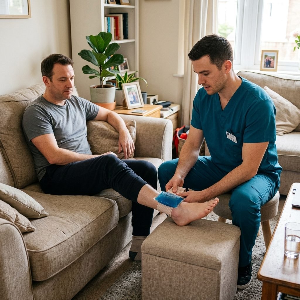
Orthopedic physical therapy is a specialized branch of rehabilitation dedicated to evaluating and treating injuries and disorders of the musculoskeletal system, including muscles, bones, joints, ligaments, tendons, and cartilage. Following orthopedic surgery (such as joint replacements or ligament reconstructions), fractures, or chronic degenerative conditions like osteoarthritis, the affected tissues require structured rehabilitation to heal properly and regain function. Our home-based clinical orthopedic rehabilitation programs are designed around the biology of tissue healing. By applying progressive mechanical loading, manual therapy, and specialized exercise protocols, we guide tissues through the inflammatory, proliferative, and remodeling phases. This structured home-based clinical approach is essential to restore joint biomechanics, rebuild muscle strength, manage pain, and facilitate a safe return to normal functional mobility.
Pathophysiology of Musculoskeletal Injuries and Tissue Healing Phases
Musculoskeletal injuries, whether acute trauma (like ligament sprains and tendon tears) or chronic degeneration (like osteoarthritis), trigger a cascade of tissue responses. Osteoarthritis is characterized by progressive degradation of articular cartilage, subchondral bone remodeling, and osteophyte formation, leading to mechanical pain, joint space narrowing, and range of motion loss. In contrast, acute soft tissue injuries undergo a cellular response that progresses through three distinct phases: the inflammatory phase, the proliferative phase, and the remodeling phase.
The inflammatory phase begins immediately after injury and lasts 3 to 5 days, characterized by vasodilation, edema, and neutrophil infiltration to clear cellular debris. The proliferative phase occurs from day 4 to day 21, during which fibroblasts lay down disorganized Type III collagen, forming immature scar tissue. The remodeling phase begins around week 3 and can last for over a year. During this final phase, the disorganized Type III collagen is gradually replaced by stronger Type I collagen, which aligns along lines of mechanical stress to restore tensile strength.
Orthopedic physical therapy utilizes controlled mechanical loading to optimize this healing process. If tissues are immobilized for too long, the collagen fibers remodel in disorganized patterns, leading to weak tissue and adhesions. Conversely, excessive early loading can disrupt healing tissue and cause chronic inflammation. Our therapists apply progressive resistance and mobilization exercises designed to match tissue tolerance, promoting optimal collagen alignment and preventing scar tissue formation. This biological approach ensures that the healed tissue can withstand functional demands.
Clinical Assessment Protocols for Musculoskeletal Deficits
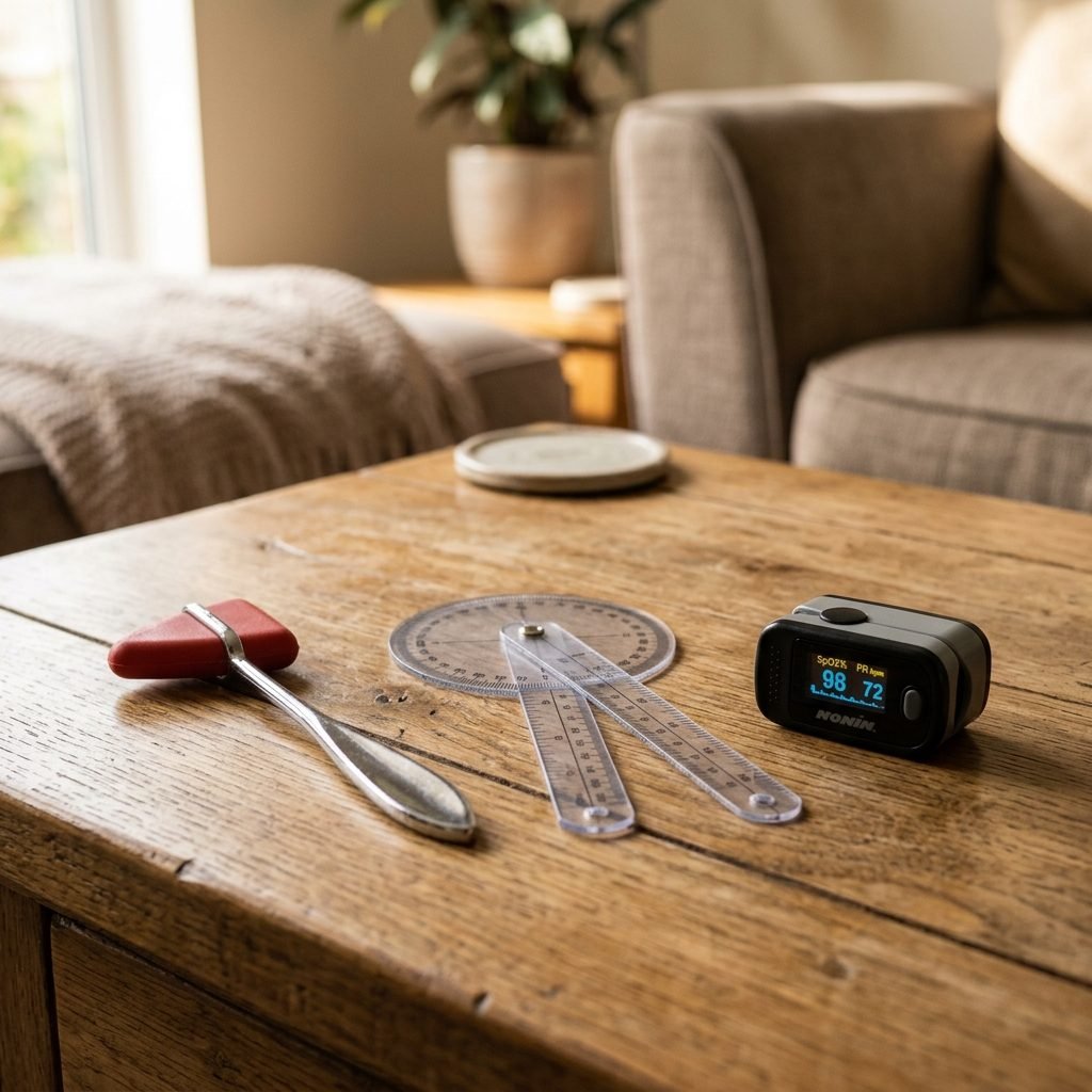
An accurate diagnostic assessment is essential to identify the root cause of orthopedic deficits and design a targeted rehabilitation plan. Our physical therapists conduct a comprehensive evaluation, starting with joint range of motion (ROM) measurements using goniometry and manual muscle testing (MMT) to assess strength. We perform palpation to locate tenderness and evaluate joint effusion (swelling) using tests like the Stroke Test or Balloon Sign for the knee, grading effusion from 0 to 3+.
We perform specialized clinical orthopedic tests to evaluate structural integrity. For knee assessments, we use the Lachman Test and Anterior Drawer Test to evaluate anterior cruciate ligament (ACL) integrity, and the McMurray Test to screen for meniscal tears. For shoulder pathology, we utilize the Hawkins-Kennedy and Neer Tests to evaluate subacromial impingement, and the Empty Can Test to assess supraspinatus tendon pathology. These specialized tests allow us to isolate specific structures, identify tears or impingements, and formulate targeted clinical diagnoses.
In addition to structural tests, we analyze posture and gait to identify biomechanical compensations. Patients with lower extremity pathology often present with an antalgic (pain-avoidant) gait, characterized by a shortened stance phase on the affected limb. We assess for abnormalities like pelvic drops, knee hyperextension, or foot pronation, which can cause secondary strain on the spine or adjacent joints. By identifying these biomechanical compensations during our initial 60-minute assessment, we can design corrective exercises that restore proper movement patterns and prevent secondary pain.
Phase 1: Acute Protection and Pain Management
During the acute phase of recovery, or immediately following orthopedic surgery, physical therapy focuses on protecting healing tissues, managing pain, and reducing inflammatory swelling. We implement the clinical POLICE principle (Protect, Optimal Loading, Ice, Compression, Elevation) to guide early management. Protection may involve assistive devices or braces to shield the joint, while optimal loading introduces gentle, pain-free movements (such as ankle pumps or setting exercises) to prevent muscle inhibition and improve blood flow without straining healing tissues.
Pain and edema are managed using targeted therapeutic modalities. We utilize transcutaneous electrical nerve stimulation (TENS) to manage acute pain, sending electrical currents to stimulate sensory nerves and block pain signals from reaching the brain (gate control theory). Localized cryotherapy (ice therapy) is applied for 15 to 20 minutes to constrict blood vessels, reduce swelling, and numb nerve endings. Therapists perform gentle passive range of motion exercises (PROM) within pain-free limits to maintain joint mobility and promote synovial fluid circulation.
Patient education is prioritized to ensure compliance with post-operative precautions and weight-bearing restrictions. For patients recovering from total hip replacement, we instruct them on hip precautions, such as avoiding hip flexion beyond 90 degrees, adduction past midline, and internal rotation to prevent joint dislocation. We instruct caregivers on safe transferring techniques, ensuring the patient's home environment is prepared to prevent falls. This early protection establishes a safe foundation for active rehabilitation.
Phase 2: Subacute Mobilization and Progressive Loading
Once the acute inflammatory phase resolves and tissue healing enters the proliferative stage, the rehabilitation program progresses to subacute mobilization. The focus shifts to restoring full joint range of motion, improving tissue flexibility, and initiating active strengthening. We transition from passive to active-assisted (AAROM) and active (AROM) movements, utilizing stretching techniques to target shortened muscles and tight joint capsules. Gentle joint mobilizations (Maitland Grade I and II) are applied to reduce pain and improve accessory joint motion.
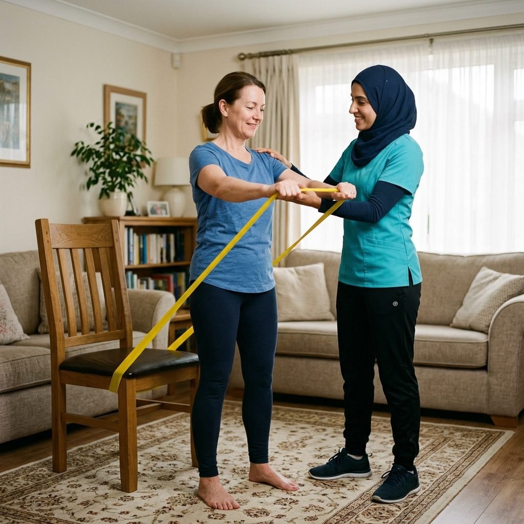
Progressive strengthening is initiated using open kinetic chain (OKC) and closed kinetic chain (CKC) exercises. Open kinetic chain exercises, where the distal segment of the limb moves freely (such as seated knee extensions), are used to isolate specific muscles. Closed kinetic chain exercises, where the foot or hand remains fixed on a surface (such as squats or leg presses), are introduced to engage multiple joints and muscles, improving joint stability and functional capacity. We apply progressive resistance training, starting with light loads (elastic bands or light weights) and progressing as tissue tolerance improves.
We implement specialized tendon loading protocols for patients recovering from chronic tendinopathies (such as Achilles or patellar tendonitis). We utilize the Alfredson Eccentric Training Protocol, which involves performing slow, controlled eccentric (lengthening) contractions under load. This specific mechanical strain stimulates tenocyte activity, promoting tendon remodeling and collagen synthesis. Therapists monitor pain levels during loading, ensuring that pain remains within acceptable limits and does not persist into the following day, which would indicate overtraining.
Phase 3: Advanced Strengthening and Functional Return
The final phase of orthopedic physical therapy focuses on advanced strengthening, functional retraining, and preparing the patient to return to work or sports activities. We transition to high-intensity resistance training, multi-directional agility drills, and sport-specific movements. Exercises are designed to challenge the musculoskeletal system in multiple planes of motion (sagittal, frontal, and transverse), improving structural integrity and dynamic stability under loaded conditions.
We integrate advanced proprioceptive and balance training to restore neuromuscular control. Patients perform exercises on unstable surfaces (such as balance boards or foam pads), single-leg stability drills, and reaction-time challenges. This training improves the speed and coordination of stabilizing muscle contractions, reducing the risk of reinjury. We utilize plyometric exercises, such as jump-landing training, to rebuild explosive power and teach safe landing mechanics, focusing on preventing dynamic knee valgus (inward collapsing of the knee), which is a major risk factor for ACL injuries.
As the patient approaches their functional goals, we design a comprehensive, long-term home exercise program. This program details specific exercises, sets, repetitions, and progression guidelines to maintain structural strength and prevent future musculoskeletal issues. Our physical therapists conduct a final functional assessment to ensure the patient meets all clinical criteria for a safe return to full activity. This ensures that the recovery achieved during active physical therapy is sustained long-term, preserving joint health and functional independence.
Long-Term Musculoskeletal Maintenance and Joint Care
Maintaining musculoskeletal health and preventing degenerative changes requires long-term commitment to exercise and joint care. Once clinical physical therapy has concluded, patients must continue home-based protocols to prevent muscle imbalances and maintain joint range of motion. This is especially important for patients with chronic conditions like osteoarthritis or those who have undergone major surgeries. Our team designs personalized maintenance programs focused on preserving joint health and structural strength.
We emphasize joint preservation strategies to minimize mechanical wear and tear on weight-bearing joints. This includes education on body weight management to reduce joint loading, using supportive footwear, and modifying daily activities to protect joints. We recommend low-impact exercises, such as swimming, cycling, or brisk walking, to promote joint mobility and cardiovascular health without overloading the joints. Our therapists provide guidance on ergonomics, helping patients set up home and work spaces to prevent repetitive strain and joint overload.
Periodic follow-up evaluations are scheduled to monitor joint health and functional status. During these check-ins, the physical therapist assesses joint range of motion, muscle strength, and movement mechanics to identify any emerging deficits. If early signs of degeneration or muscle weakness are detected, we modify the home program or schedule brief clinical sessions to address the issues proactively. This ongoing support ensures that the gains made during rehabilitation are preserved, protecting long-term musculoskeletal health.
Clinical Precautions, Contraindications, and Safety Standards
Patient safety is the top priority in orthopedic physical therapy, requiring strict adherence to clinical protocols and contraindications. Absolute contraindications to physical therapy include unhealed fractures, acute deep vein thrombosis (DVT), acute local infection (such as osteomyelitis or septic arthritis), and severe cardiovascular instability. Initiating therapy under these conditions can cause severe complications, such as fracture displacement, pulmonary embolism, or systemic infection.
Relative contraindications require caution and modification of exercise protocols. These include severe osteoporosis, joint hypermobility, and the presence of surgical hardware with specific movement restrictions. For patients with osteoporosis, we avoid high-impact activities, explosive twisting movements, and loaded spinal flexion to prevent pathological fractures. For post-surgical patients, we strictly adhere to surgeon-specified restrictions, such as weight-bearing status (e.g., non-weight bearing, partial weight bearing, or weight bearing as tolerated) and range of motion limits.
Therapists monitor for clinical red flags during every session, such as sudden increases in localized pain, swelling, warmth, redness, or a sudden loss of joint mobility. The presence of calf pain, warmth, and swelling is a red flag for DVT, requiring immediate medical referral. We monitor the patient's physiological response to exercise, ensuring that pain does not exceed a mild discomfort during stretching and strengthening. By maintaining these strict safety standards, we ensure a safe, effective recovery process.
Unified Home Physical Therapy Standards & Geographical Coverage
Bidayah Center delivers highly structured home rehabilitation and clinical conditioning across all served territories, including Jeddah, Makkah, and Qatif districts. We deploy licensed physical therapists directly to your home, ensuring that you receive the same standards of care, specialized portable modalities, and treatment protocols as premier inpatient facilities.
Our medical coordinators screen each referral and coordinate field operations using regional dispatch networks to ensure prompt scheduling and strict adherence to appointment times. We maintain active communication channels with orthopedic surgeons and neurologists at major local hospitals to coordinate care.
While this generic page outlines the core clinical details of this service, we invite you to choose your specific city page. Doing so allows you to explore local neighborhood guidelines, read region-specific FAQs, and coordinate with male or female physical therapists according to the clinical needs of your family.
📚Clinically Verified Sources & References
- [MOH-01]Guide to Home Healthcare Services — Saudi Ministry of Health (2023). View Scientific Source
- [WHO-01]Rehabilitation for stroke patients — World Health Organization (2022). View Scientific Source