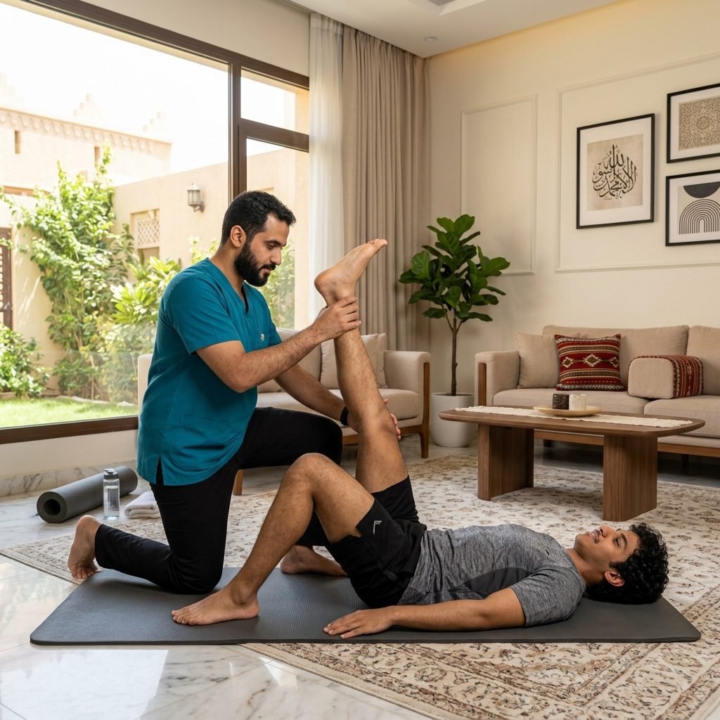
Restoring joint range of motion (ROM) is a vital milestone in musculoskeletal and neuromuscular rehabilitation. Following joint injury, prolonged immobilization, or surgical intervention, soft tissues undergo rapid biological changes, including collagen cross-linking, synovial fluid reduction, and capsular contractures. If left untreated, these alterations lead to permanent joint stiffness, structural contractures, and compensatory gait or movement abnormalities that accelerate degenerative joint disease. Our home-based clinical ROM restoration programs focus on reversing these processes through targeted, phase-specific interventions. By combining gentle manual joint mobilization, active and passive stretching, and neuromuscular facilitation techniques, we restore normal joint sliding and rolling mechanics. This clinical approach is designed to eliminate functional restrictions, alleviate mechanical pain, and maximize independence in daily activities within the patient's domestic setting.
Pathophysiology of Joint Stiffness and Tissue Contractures
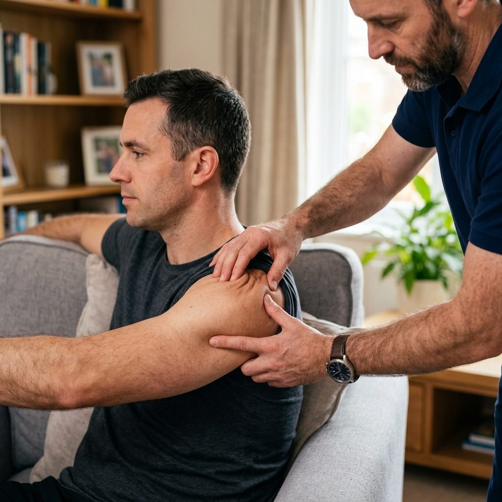
Joint stiffness and range of motion restriction result from complex pathophysiological changes within articular and periarticular structures. When a joint is immobilized, synovial fluid production decreases, reducing joint lubrication and causing cartilage malnutrition. At the same time, the surrounding connective tissue undergoes morphological remodeling. Collagen fibers, which normally slide past one another, form abnormal cross-links, transforming the joint capsule and ligaments into dense, unyielding fibrotic tissue. This process, known as capsular contracture, significantly limits the joint's ability to undergo normal translation and rotation.
In addition to capsular changes, skeletal muscles and tendons surrounding the joint undergo adaptive shortening. Under chronic disuse or neural spasticity, sarcomeres—the functional contractile units of muscle fibers—decrease in number, leading to myogenic contractures. Tendons lose their elastic properties, and fascial sheets become bound by myofascial adhesions. This combination of muscle tightness and joint capsule thickening restricts both active range of motion (AROM, controlled by the patient's muscles) and passive range of motion (PROM, achieved via external assistance), leading to severe functional impairment.
Clinical range of motion restoration aims to stimulate tissue remodeling by applying controlled mechanical tension. Consistent, progressive stretching triggers a process called mechanotransduction, where cellular signals stimulate fibroblast activity. This activity promotes the deposition of new collagen fibers in parallel alignments and degrades abnormal cross-links through matrix metalloproteinase enzymes. Furthermore, prolonged low-load stretching facilitates plastic deformation of connective tissue, allowing the joint structures to safely elongate and maintain their restored lengths over time.
Clinical Assessment of Range of Motion and Joint Biomechanics
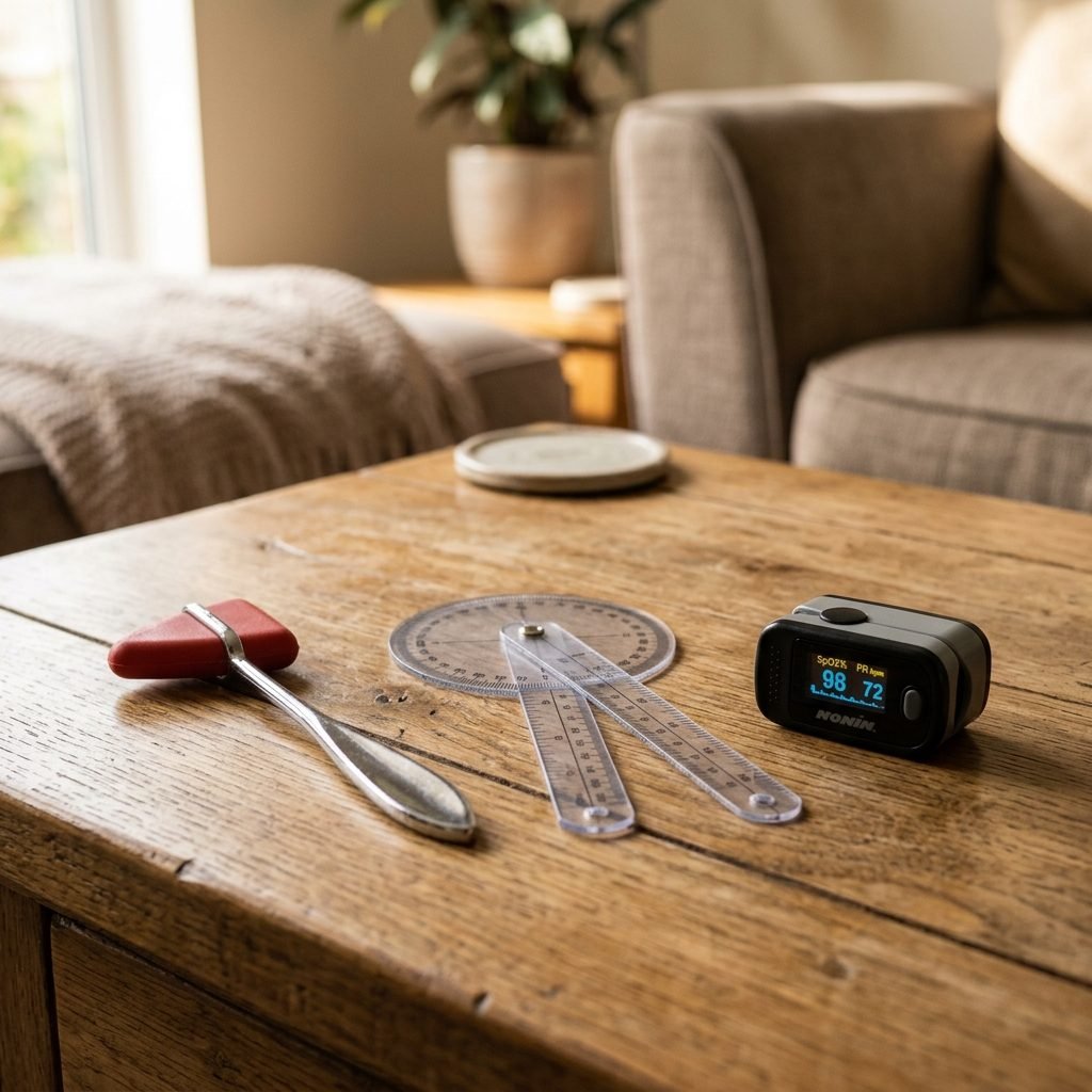
A precise clinical assessment is essential to determine the causes of joint restriction and guide physical therapy. Our therapists begin by evaluating active and passive range of motion using a clinical goniometer. We measure specific joint angles in degrees at the onset of pain and at the limit of movement. Comparing active range of motion to passive range of motion allows us to differentiate between muscular weakness (where passive ROM is greater than active ROM) and joint capsule or structural restrictions (where both active and passive ROM are equally limited).
During passive range of motion assessment, the therapist evaluates the 'end-feel'—the sensation felt at the extreme limit of joint movement. A normal end-feel can be soft (tissue approximation, like knee flexion), firm (capsular or ligamentous stretch), or hard (bone-to-bone contact, like elbow extension). Pathological end-feels, such as an empty end-feel (movement stopped due to severe pain before mechanical resistance), a springy block (internal joint derangement like a meniscus tear), or a premature hard end-feel (osteophytes or heterotopic ossification), provide critical diagnostic information that determines treatment viability.
Additionally, we conduct specific muscle length testing and assess accessory joint motion, such as arthrokinematic glide and roll. Muscles that cross two joints, such as the hamstrings or rectus femoris, are tested using specialized tests like the Thomas Test or 90-90 Straight Leg Raise. We monitor the patient's pain levels, looking for patterns like pain before resistance (acute inflammation), pain at resistance (subacute), or pain after resistance (chronic tissue restriction). These parameters help us design a personalized, safe mobilization protocol that avoids tissue damage.
Phase 1: Early Mobilization and Passive Range of Motion (PROM)
In the acute phase post-injury or post-surgery, when active muscle contraction could disrupt healing tissues, passive range of motion (PROM) is the primary therapeutic intervention. During PROM, the physical therapist supports the limb and moves the joint through a pain-free range without any effort from the patient. This helps maintain joint mobility, stimulates synovial fluid circulation to nourish articular cartilage, prevents venous stasis, and decreases the formation of intra-articular adhesions. PROM also helps inhibit pain pathways through sensory stimulation of joint mechanoreceptors.
To enhance tissue extensibility, our therapists may apply localized thermal modalities, such as moist heat packs, prior to mobilization. Superficial heat increases local blood flow, relaxes surrounding muscles, and increases collagen tissue viscoelasticity, making the subsequent stretching safer and more effective. For joints with severe restrictions, gentle joint mobilization techniques (Maitland Grade I and II oscillations) are applied. These low-amplitude movements work within the pain-free range to reduce joint pain, manage muscle guarding, and prepare the joint for deeper tissue stretching.
Patient safety is maintained through continuous feedback and monitoring. The therapist moves the joint slowly and rhythmically, avoiding sudden jerks that could trigger muscle guarding or damage healing tissues. Caregivers are educated on basic positioning techniques, splinting compliance, and safe support methods to ensure the joint is protected between therapy sessions. This collaborative approach ensures that early mobilization goals are achieved safely, paving the way for active-assisted exercises in the next phase.
Phase 2: Active-Assisted and Active Stretching Protocols
As tissue healing progresses and the patient is cleared to actively contract muscles, the therapy shifts to Active-Assisted Range of Motion (AAROM). During AAROM, the patient initiates joint movement, and the therapist or an assistive device (such as a strap, pulley, or cane) provides the support needed to complete the motion. This phase serves as a bridge to independent movement, beginning to rebuild muscle strength while continuing to expand joint range. It helps retrain the nervous system to coordinate joint movement, reducing muscular inhibition caused by pain.
Once AAROM is tolerated, the patient progresses to Active Range of Motion (AROM) and targeted stretching. We utilize Proprioceptive Neuromuscular Facilitation (PNF) stretching techniques, particularly the 'Contract-Relax' and 'Hold-Relax' protocols. These techniques utilize neurological inhibition pathways to relax hyperactive muscles. For example, during a Hold-Relax stretch of the hamstrings, the patient performs an isometric contraction against the therapist's resistance, followed by voluntary relaxation, during which the therapist gently moves the limb into a deeper passive stretch. This utilizes the autogenic inhibition reflex mediated by Golgi tendon organs to achieve rapid increases in muscle length.
In addition to PNF, static stretching is prescribed to target chronic connective tissue tightness. Patients are instructed to hold stretches for 30 to 60 seconds at a point of mild discomfort, repeating the stretch 3 to 4 times per set. Dynamic stretching, involving controlled movements through the joint's functional range, is introduced to prepare the musculoskeletal system for daily activities. The therapist constantly monitors movement quality to prevent compensatory patterns, such as shrugging the shoulder to compensate for limited glenohumeral elevation.
Phase 3: Advanced Mobilization and Functional Integration
The final phase of ROM rehabilitation focuses on restoring full accessory joint motion and integrating range of motion into functional movement patterns. Our therapists apply advanced manual therapy techniques, including Maitland Grade III and IV joint mobilizations. Unlike earlier grades, Grade III and IV mobilizations involve large and small-amplitude oscillations pushed into the point of tissue resistance. These techniques are designed to stretch tight joint capsules and break down deep adhesions, restoring the natural glide and roll mechanics required for pain-free joint function.
To ensure that restored mobility is maintained long-term, it must be integrated into active functional movements. Muscles must be strengthened in their newly acquired ranges of motion to establish neuromuscular control and joint stability. If a joint gains range but lacks the muscular strength to control that range, it becomes susceptible to instability and reinjury. We prescribe progressive eccentric strengthening exercises, functional reaching tasks, and multi-joint kinetic chain movements to ensure that the patient can safely utilize their restored mobility during daily activities.
At this stage, we also address ergonomics and body mechanics. The therapist analyzes the patient's daily routines and home environment to identify habits that may contribute to joint stiffness. For example, a patient recovering from a frozen shoulder will be guided on correct sleeping posture, workspace setup, and home exercises. A comprehensive, customized home exercise plan is provided, detailing specific stretching and strengthening exercises to preserve range of motion and prevent regression, ensuring lasting physical independence.
Long-Term Compliance and Joint Preservation Strategies
Maintaining joint flexibility and preserving cartilage health requires continuous long-term compliance with stretching and movement protocols. Once clinical physical therapy has concluded, the joint structures can begin to contract if they are not regularly moved through their full ranges. This is particularly true for patients with chronic conditions like osteoarthritis, rheumatoid arthritis, or post-surgical scarring. Our therapists design a structured, realistic maintenance program that fits into the patient's daily routine, emphasizing joint preservation techniques and regular active stretching.
Joint preservation strategies are taught to minimize cumulative mechanical stress on weight-bearing joints. This includes education on body weight management to reduce joint loading, utilizing assistive devices when necessary, and avoiding prolonged static postures that lead to joint stiffness. We encourage low-impact cardiovascular exercises, such as swimming or stationary cycling, which promote synovial fluid circulation and cartilage health without overloading the joints. Patients are instructed on how to modify activities of daily living (ADLs) to protect compromised joints.
Follow-up assessments are scheduled periodically to monitor joint range of motion and functional status. During these check-ins, the physical therapist re-evaluates goniometric measurements and assesses for any signs of regression or tissue tightening. If early signs of stiffness are detected, the home stretching program is modified, or brief clinical sessions are scheduled to apply targeted manual mobilizations. This proactive approach ensures that the gains made during rehabilitation are preserved, protecting joint health and maintaining the patient's quality of life.
Clinical Precautions, Contraindications, and Safety Red Flags
Ensuring patient safety during joint mobilization and stretching requires strict adherence to clinical guidelines and contraindications. Absolute contraindications to range of motion restoration exercises include unhealed fractures, acute joint infection (infectious arthritis), acute local inflammation, joint subluxation or dislocation, and suspected deep vein thrombosis (DVT) in the affected limb. Performing stretches under these conditions can cause severe tissue damage, exacerbate inflammation, or dislodge thrombi, leading to life-threatening complications.
Relative contraindications require caution and modification of treatment protocols. These include severe osteoporosis, joint hypermobility syndromes, active rheumatoid arthritis flare-ups, and the presence of hematomas. In patients with osteoporosis, high-velocity mobilizations and intense passive stretching are avoided to prevent pathological bone fractures. For patients with joint hypermobility, therapy focuses on muscular stabilization and neuromuscular control rather than expanding range of motion, keeping movement within normal physiological limits.
Therapists monitor for clinical red flags during every session, such as sudden increases in localized pain, swelling, warmth, or a sudden loss of range of motion. An unexpected decrease in ROM accompanied by sharp pain often indicates underlying tissue damage or mechanical joint locking, requiring immediate cessation of therapy and medical evaluation. We emphasize the importance of pacing, ensuring that stretching is performed to the point of mild tension, never pain, to prevent micro-tears in the muscle-tendon unit and subsequent scar tissue formation.
Unified Home Physical Therapy Standards & Geographical Coverage
Bidayah Center delivers highly structured home rehabilitation and clinical conditioning across all served territories, including Jeddah, Makkah, and Qatif districts. We deploy licensed physical therapists directly to your home, ensuring that you receive the same standards of care, specialized portable modalities, and treatment protocols as premier inpatient facilities.
Our medical coordinators screen each referral and coordinate field operations using regional dispatch networks to ensure prompt scheduling and strict adherence to appointment times. We maintain active communication channels with orthopedic surgeons and neurologists at major local hospitals to coordinate care.
While this generic page outlines the core clinical details of this service, we invite you to choose your specific city page. Doing so allows you to explore local neighborhood guidelines, read region-specific FAQs, and coordinate with male or female physical therapists according to the clinical needs of your family.
📚Clinically Verified Sources & References
- [MOH-01]Guide to Home Healthcare Services — Saudi Ministry of Health (2023). View Scientific Source
- [WHO-01]Rehabilitation for stroke patients — World Health Organization (2022). View Scientific Source